Anatomical Drawing Of A Heart
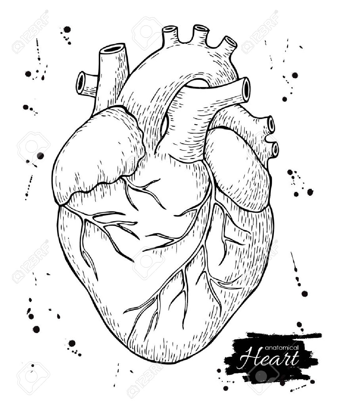
Anatomical Drawing Of A Heart - Human organs hand drawn line icon set. We will then proceed to shape the heart, slowly refining it with our pencils into a more realistic heart drawing. The right margin is the small section of the right atrium that extends between the superior and inferior vena cava. Web heart drawing realistic anatomical beauty: Search by image or video. The heart has five surfaces: Web anyway, in this assortment you can take a look at the anatomy of the human heart. Then, fill in the base of the heart with the right and left ventricles and the right and left atriums. These anatomical heart medical illustrations are highly detailed drawings that blend art with science. It should look a bit like the shape of africa.
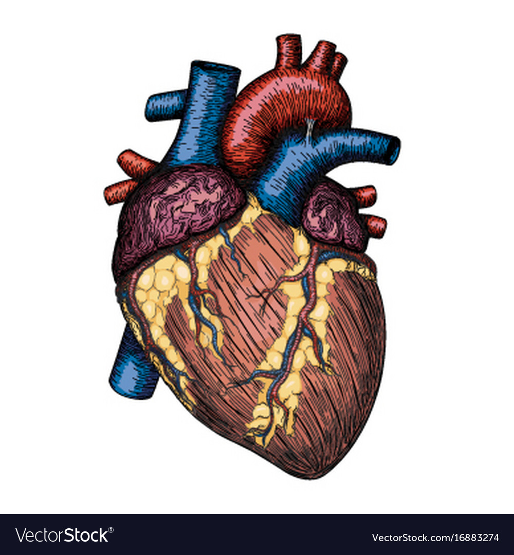
A thin layer of tissue, the pericardium, covers the outside, and another layer, the endocardium, lines the inside. The average human heart weighs between. Pencil sketch of the human heart embark on a journey into the beauty of human anatomy with a mesmerizing pencil sketch of the human heart. Draw the main shape of your human heart drawing. The heart has five surfaces: Begin by sketching a rounded, lumpy, irregular figure. The atria act as receiving chambers for blood, so they are connected to the veins that carry blood to the heart. Web the heart contains 4 chambers: Angle the slightly tampered end of the shape to the left about 120 degrees. The right margin is the small section of the right atrium that extends between the superior and inferior vena cava.
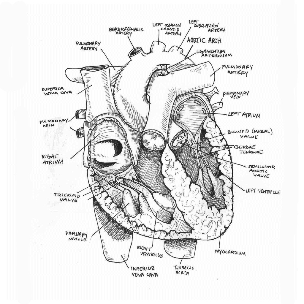
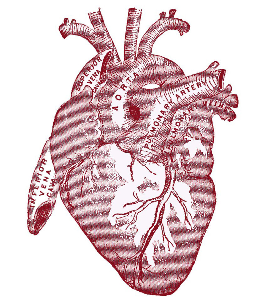
Web to draw the internal structure of the heart, start by sketching the 2 pulmonary veins to the lower left of the aorta and the bottom of the inferior vena cava slightly to the right of that. Xxxl very detailed human heart. Base (posterior), diaphragmatic (inferior), sternocostal (anterior), and left and right pulmonary surfaces. Web the heart is divided into four chambers: View anatomical heart drawing videos. Web this interactive atlas of human heart anatomy is based on medical illustrations and cadaver photography. Included below are a magnificent color heart illustration, along with four monotype prints, which are possibly woodcuts, engravings, or lithographs. How to draw body parts. Angle the slightly tampered end of the shape to the left about 120 degrees. Draw the first construction lines.
Anatomical Drawing Heart at GetDrawings Free download
Within the triangle, draw a horizontal and vertical centerline to split the triangle into four pieces. Web browse 873 heart anatomy drawing photos and images available, or start a new search to explore more photos and images. Human organs hand drawn line icon set. The heart cavity is divided down the middle into a right and a left heart, which.
Human heart hand drawn anatomical sketch Vector Image
[1] the main shape will be the basis for the left and right ventricles. Xxxl very detailed human heart. Blood is transported through the body via a complex network of veins and arteries. Human organs hand drawn line icon set. Included below are a magnificent color heart illustration, along with four monotype prints, which are possibly woodcuts, engravings, or lithographs.

Labelled Heart
Anatomical heart drawing stock photos are available in a variety of sizes and formats to fit your needs. Draw the first construction lines. Web browse 947 heart anatomical drawing photos and images available, or start a new search to explore more photos and images. The right margin is the small section of the right atrium that extends between the superior.
Anatomical Drawing Heart at GetDrawings Free download
Base (posterior), diaphragmatic (inferior), sternocostal (anterior), and left and right pulmonary surfaces. Web the heart consists of several layers of a tough muscular wall, the myocardium. Draw the main shape of your human heart drawing. [1] the main shape will be the basis for the left and right ventricles. Anatomical heart drawing stock illustrations.

How to Draw the Internal Structure of the Heart (with Pictures)
Blood is transported through the body via a complex network of veins and arteries. It should look a bit like the shape of africa. In addition to reviewing the human heart anatomy, we will also discuss the function and order in which blood flows through the heart. Anatomical heart drawing stock photos are available in a variety of sizes and.
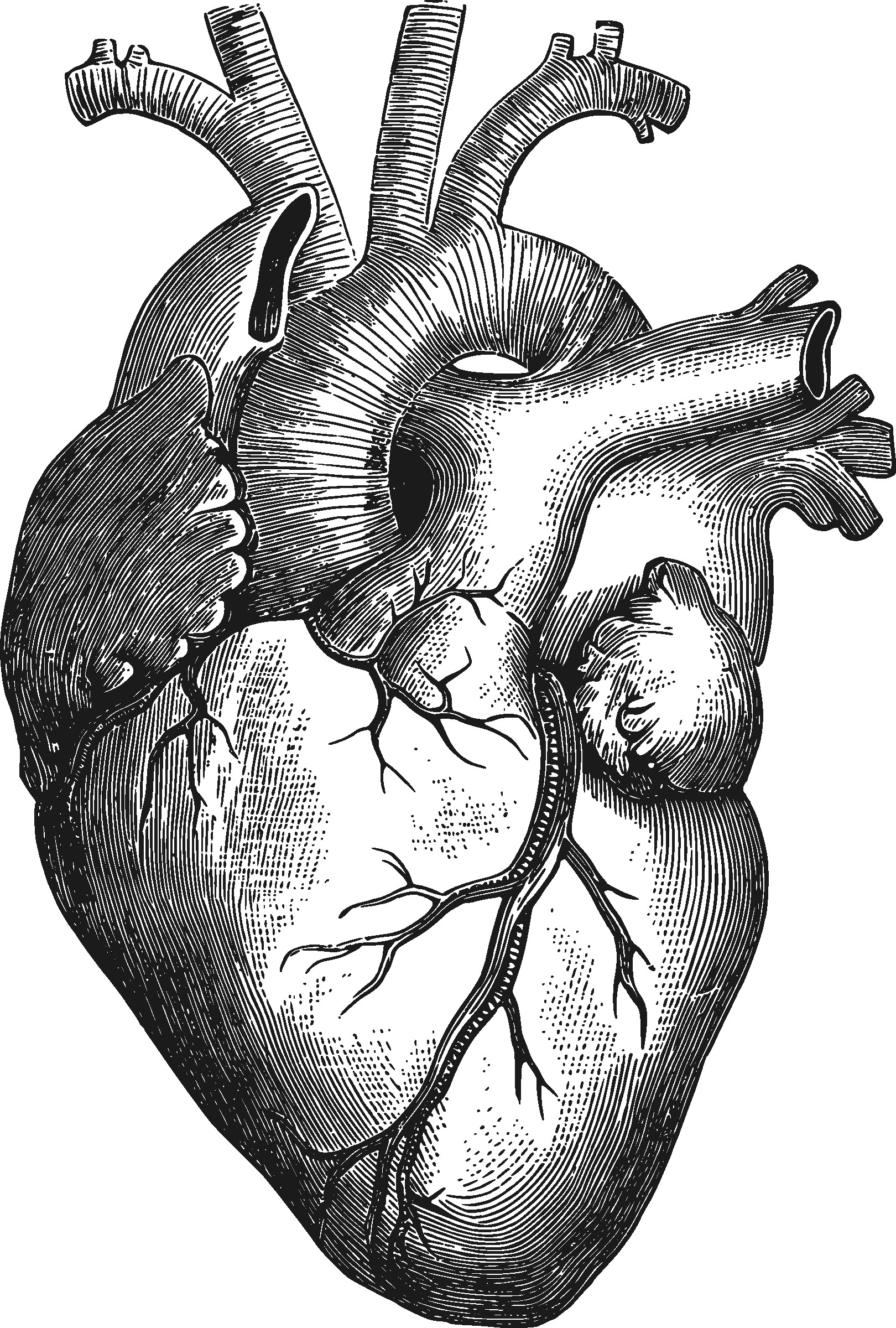
8 Anatomical Heart Drawings! The Graphics Fairy
Web create a curved shape similar to an acorn or apple’s bottom half. Web in this lecture, dr mike shows the two best ways to draw and label the heart! Web browse 873 heart anatomy drawing photos and images available, or start a new search to explore more photos and images. It should look a bit like the shape of.
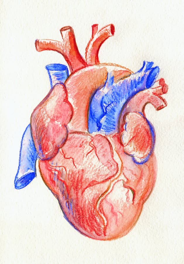
Hand Drawing Sketch Anatomical Heart. Colored Watercolor Pencil Stock
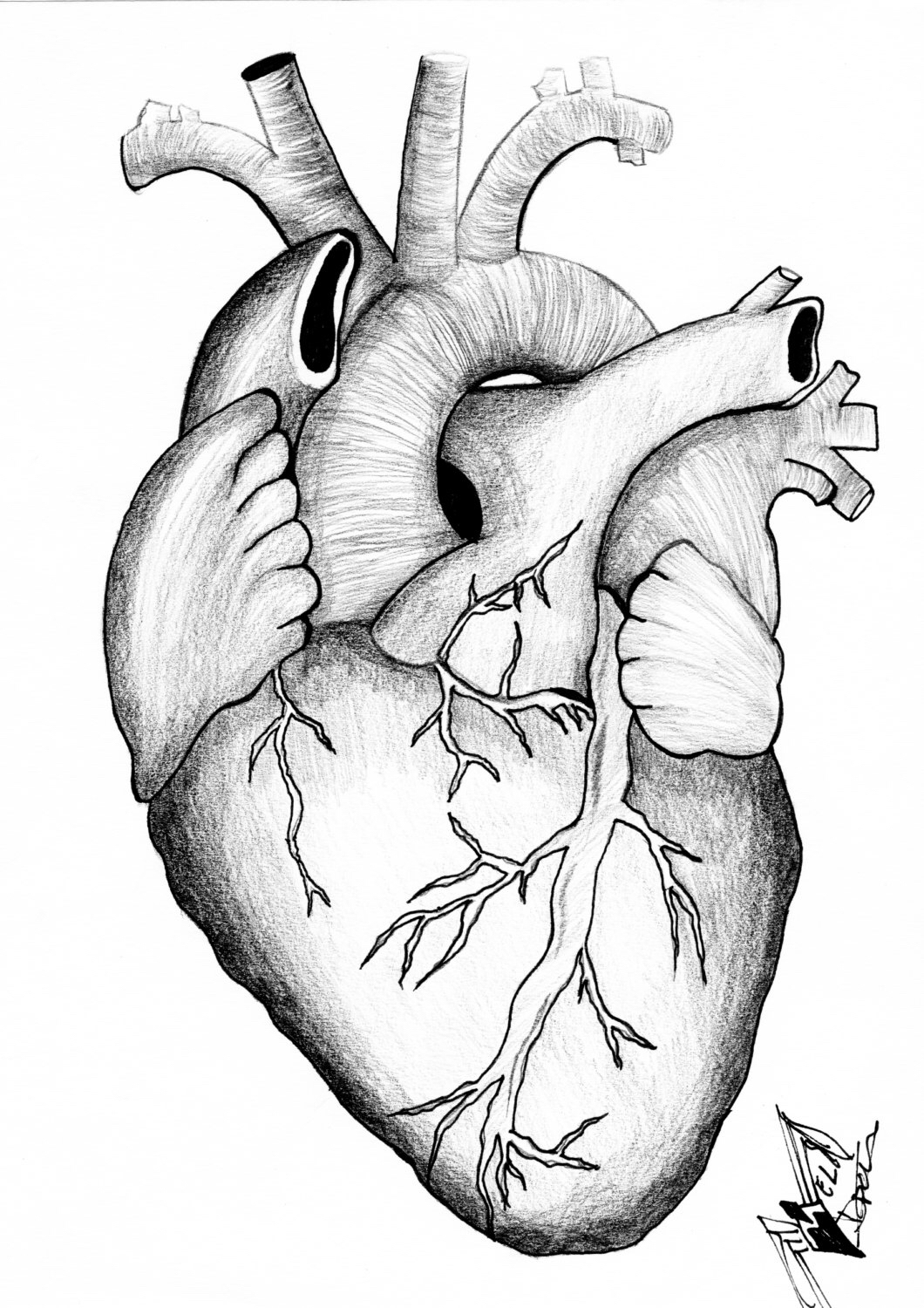
Xxxl very detailed human heart. [1] the main shape will be the basis for the left and right ventricles. Web welcome to the anatomy of the heart made easy! Pencil sketch of the human heart embark on a journey into the beauty of human anatomy with a mesmerizing pencil sketch of the human heart. Draw the first construction lines.

How to Draw a Human Heart Really Easy Drawing Tutorial
How to draw body parts. Web the heart consists of several layers of a tough muscular wall, the myocardium. Base (posterior), diaphragmatic (inferior), sternocostal (anterior), and left and right pulmonary surfaces. The atria are smaller than the ventricles and have thinner, less muscular walls than the ventricles. Anatomical heart drawing stock illustrations.
Heart Anatomy Sketch at Explore collection of
Anatomical heart drawing stock photos are available in a variety of sizes and formats to fit your needs. Human organs hand drawn line icon set. We will then proceed to shape the heart, slowly refining it with our pencils into a more realistic heart drawing. 19k views 3 years ago body part drawings: Pencil sketch of the human heart embark.
How To Draw An Anatomical Heart The ultimate guide drawboy2
Anatomical heart drawing stock photos are available in a variety of sizes and formats to fit your needs. We will then proceed to shape the heart, slowly refining it with our pencils into a more realistic heart drawing. Web this interactive atlas of human heart anatomy is based on medical illustrations and cadaver photography. Web the heart consists of several.
Human Organs Hand Drawn Line Icon Set.
Web the heart consists of several layers of a tough muscular wall, the myocardium. Search by image or video. The user can show or hide the anatomical labels which provide a useful tool to create illustrations perfectly adapted for teaching. Select from premium anatomical heart drawing images of the highest quality.
Web The Heart Contains 4 Chambers:
Web the heart is divided into four chambers: It should look a bit like the shape of africa. Human organs hand drawn line icon set. In addition to reviewing the human heart anatomy, we will also discuss the function and order in which blood flows through the heart.
Xxxl Very Detailed Human Heart.
Web to draw the internal structure of the heart, start by sketching the 2 pulmonary veins to the lower left of the aorta and the bottom of the inferior vena cava slightly to the right of that. Human organs hand drawn line icon set. The right margin is the small section of the right atrium that extends between the superior and inferior vena cava. Pencil sketch of the human heart embark on a journey into the beauty of human anatomy with a mesmerizing pencil sketch of the human heart.
19K Views 3 Years Ago Body Part Drawings:
A thin layer of tissue, the pericardium, covers the outside, and another layer, the endocardium, lines the inside. We will use labeled diagrams and pictures to learn the main cardiac structures and related vascular system. This outlines the lower chamber of the heart, which includes both the left and right ventricles. Web heart drawing realistic anatomical beauty: