Anatomical Eyeball Drawing
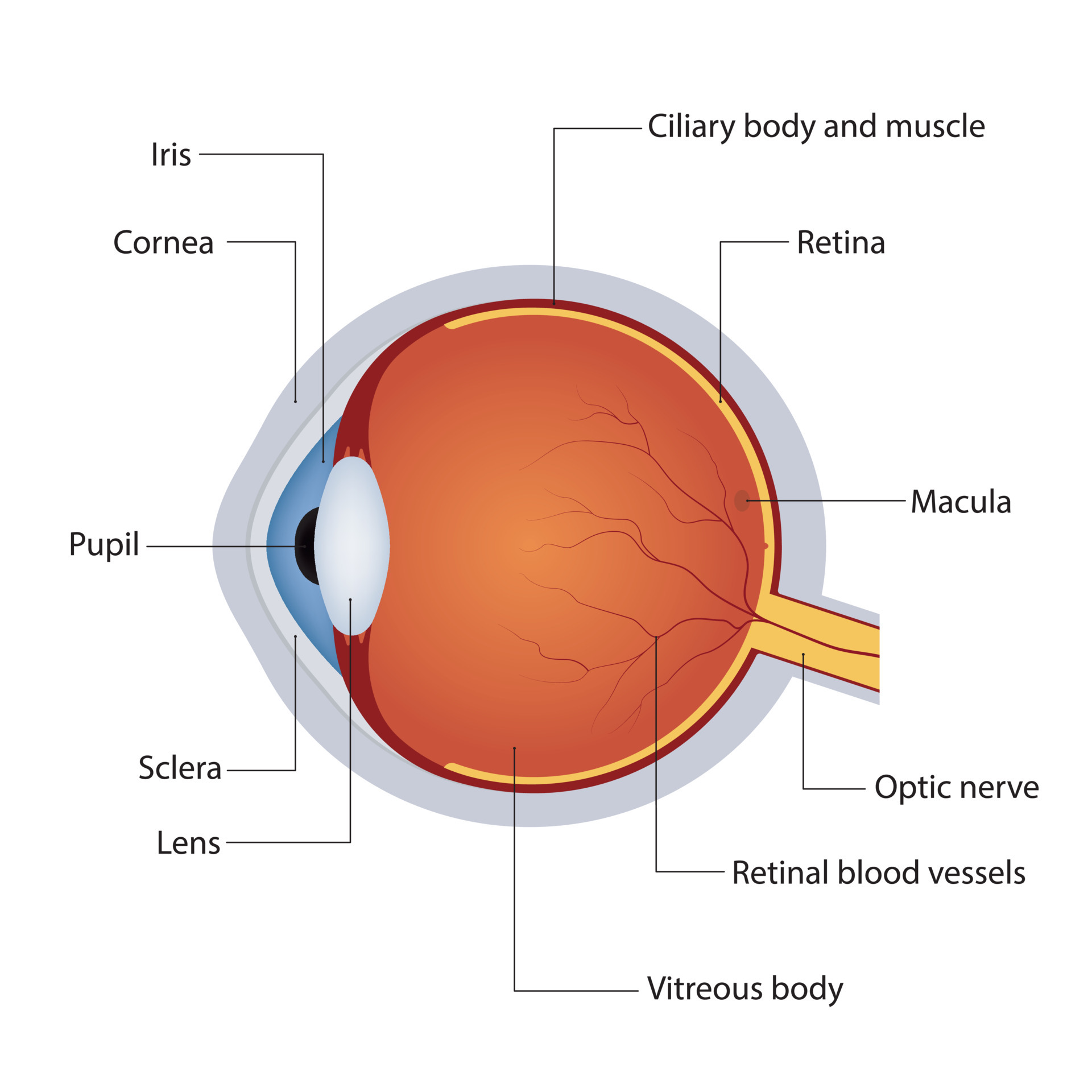
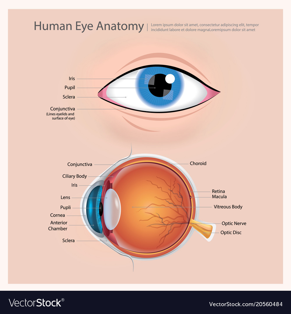
Anatomical Eyeball Drawing - The fibrous layer, which consists of the sclera and cornea. Web xl 5000x5000 jpeg included. The eyeball, eye socket, brow ridge, eyelids, tear duct, sclera, iris, pupil, cornea, glabella, and epicanthic fold. “the visual anatomical hierarchy in the macaque monkey described by felleman and van essen “shows the incomprehensible complexity of the visual system.”. Web the eyeball consists of three distinct layers. Adding in the appropriate level of detail depending on the style of. Using basic shapes to create the general silhouette of the figure. The cornea and lens bend light, the iris controls light intake, and the retina transforms light. We aim to increase the number of foreign. You will learn the basic anatomy of the eyes and study the bone structure of the eye sockets.
“the visual anatomical hierarchy in the macaque monkey described by felleman and van essen “shows the incomprehensible complexity of the visual system.”. The eyeball, eye socket, brow ridge, eyelids, tear duct, sclera, iris, pupil, cornea, glabella, and epicanthic fold. These three layers together are called the tear film. You'll find plenty of drawing resources, too, including advanced drawing techniques and links to youtube videos that others have published about the app. I will go over the structure of the eye and detailed. • 1mo • 9 min read. Steve will show you how the eyelids and brow ridges are positioned around the eyes. Science & technology 3d models. Select from premium anatomical eye drawing images of the highest quality. Web eye anatomy the first step towards drawing an eye is understanding how it functions and the individual parts that come together as a whole.
Use curved lines to show where the eyelids reveal the shape of the eye. Conjunctivitis, often known as pink eye, occurs when this thin membrane becomes inflamed or swollen. This is the most important feature of the face, and if you want to draw. The fibrous layer, which consists of the sclera and cornea. The axis of the eyes, stays the same. Web the da vinci eye app also features a handful of instructional videos to help you get to grips with the app. Web how to draw anatomy step by step. A is accommodation in diopters. The eyeball, eye socket, brow ridge, eyelids, tear duct, sclera, iris, pupil, cornea, glabella, and epicanthic fold. Justine triet's thoroughly engaging anatomy of a fall examines the way information reveals character, and vice versa, during an unfolding murder.
Human Anatomy of Eyeball 303487 Vector Art at Vecteezy
Web the ubiquity of three black athletes standing on a global sprint podium has created a dogma of biological superiority that decades of scientific evidence has so far been unable to support. Web the human eye is an organ of the sensory nervous system that reacts to visible light and allows the use of visual information for various purposes including.

Eye Anatomy
He will also show you how to place the eyes on the head. Adding in the appropriate level of detail depending on the style of. This figure is from the academy's basic ophthalmology: Web may 13, 2024 3:31pm. The eye is an organ which helps perceive light, color, and depth.
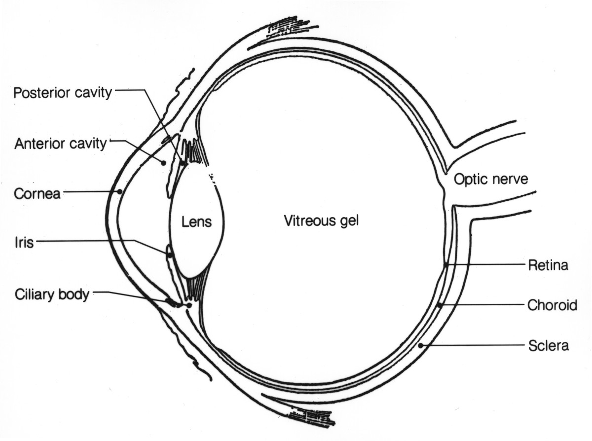
Structure of anatomy human eye. Detailed diagram of eyeball. Side view
You can simplify the process of drawing human anatomy into three general steps: • 1mo • 9 min read. Web when we're drawing the eyes at a 3/4 angle, the real about them being one eye width apart changes. Anatomical eye drawing stock photos are available in a variety of sizes. Drawing lessons in several genres, such as anime, faces,.
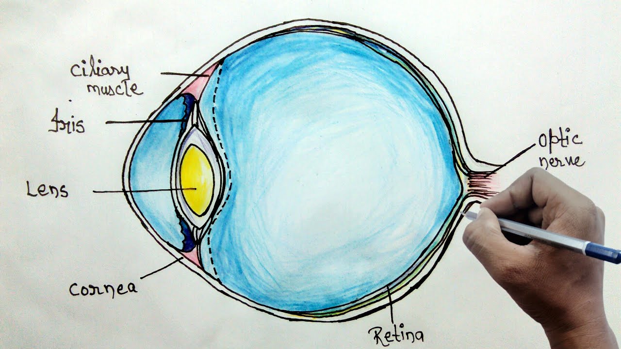
How to draw human eye diagram for beginners YouTube
All images are grouped and on separate layers making for. Felleman & van essen, 1991. When it comes to drawing realistic eyes, a solid understanding of the eye anatomy is your best friend. In this lesson, instructor steve huston will teach you how to construct the eyes. Web xl 5000x5000 jpeg included.
Anatomy of the Eye Human Eye Anatomy Owlcation
Hand drawn pencil sketches of scientific concepts. He will also show you how to place the eyes on the head. Essentials for medical students, tenth edition; This figure is from the academy's basic ophthalmology: • 1mo • 9 min read.

Anatomy of the Human Eye
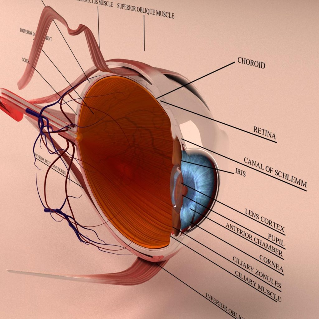
Drawing lessons in several genres, such as anime, faces, action, and. The eyeball, eye socket, brow ridge, eyelids, tear duct, sclera, iris, pupil, cornea, glabella, and epicanthic fold. Web full and extensive eye anatomy in drawing for artists. I will go over the structure of the eye and detailed. The eye can be considered as a living optical device.it is.
Human eye anatomy Royalty Free Vector Image VectorStock
The layers of the tear film keep the front of the eye lubricated. Jake borelli as levi schmitt abc. The fibrous layer, which consists of the sclera and cornea. Anatomical eye drawing stock photos are available in a variety of sizes. For more video tutorials visit www.proko.com and subscribe to the newsletter.
Anatomy Human Eye Cross Section 3D Model Kezan's Portfolio
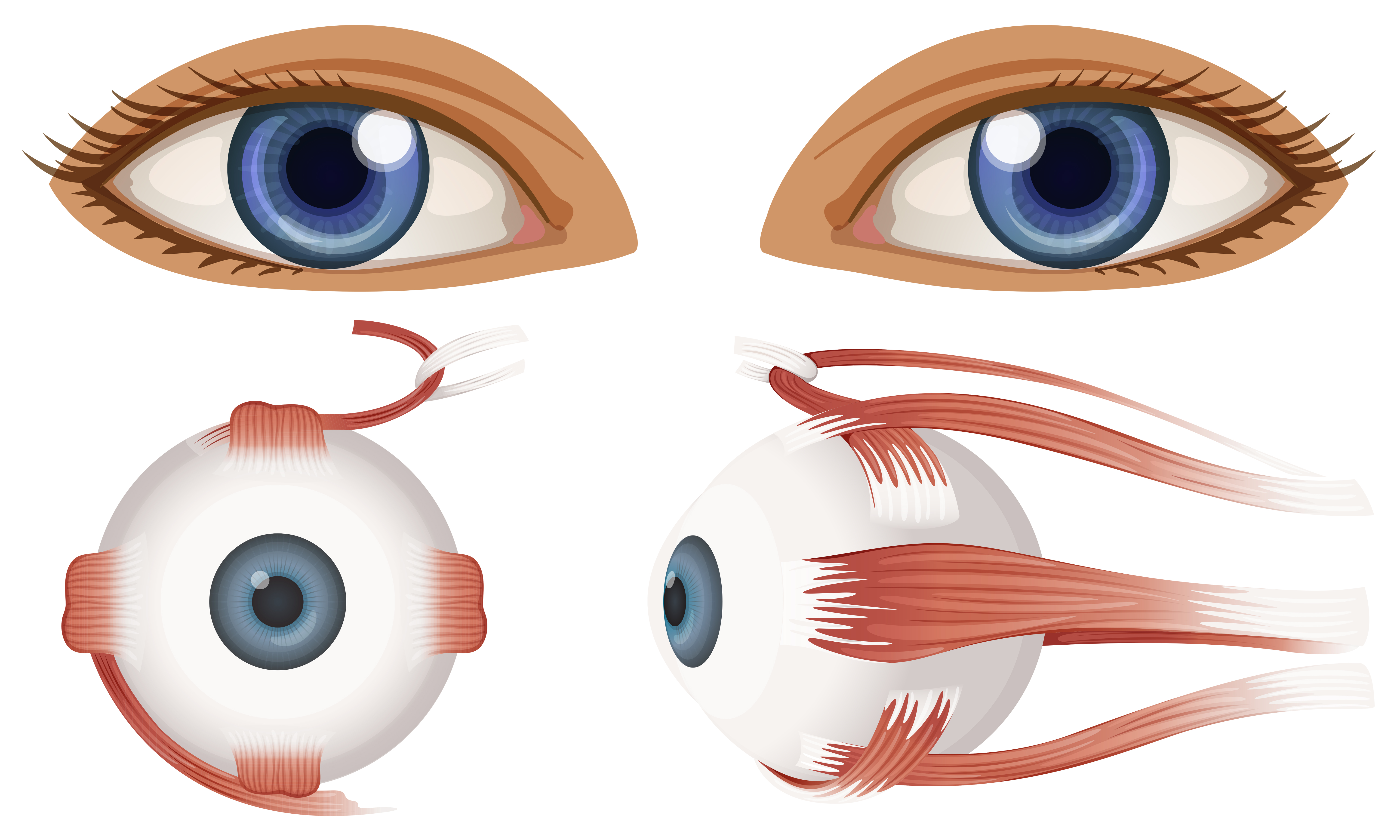
Using basic shapes to create the general silhouette of the figure. Web find anatomical eye drawing stock illustrations from getty images. External landmarks and extraocular muscles. Web xl 5000x5000 jpeg included. The cornea and lens bend light, the iris controls light intake, and the retina transforms light.
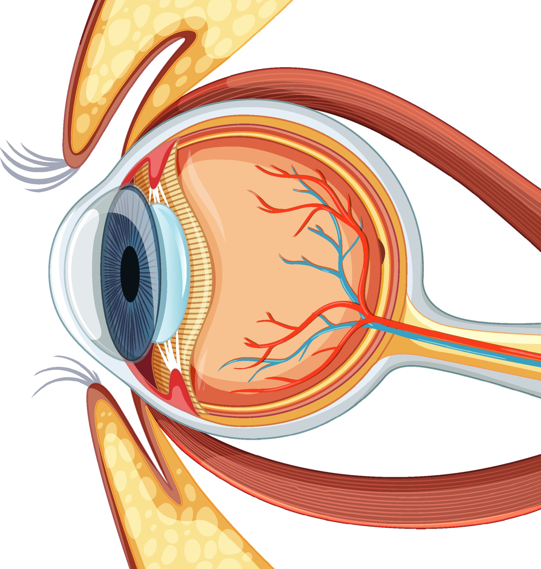
Diagram of human eyeball anatomy 3188538 Vector Art at Vecteezy
These parts work together to capture light and convert it into images. You'll find plenty of drawing resources, too, including advanced drawing techniques and links to youtube videos that others have published about the app. The eyes might be tilted at an angle, but we'll still be leveled with each other. When it comes to drawing realistic eyes, a solid.

Learn To Draw Eyes Drawing On Demand Anatomy sketches, Anatomy art
It is part of the sensory nervous system. The cornea and lens bend light, the iris controls light intake, and the retina transforms light. Tarsal plates and lacrimal system from “the human eye”. The jackpot currently sits at an. The fibrous layer, which consists of the sclera and cornea.
Hand Drawn Pencil Sketches Of Scientific Concepts.
Anatomical eye drawing stock illustrations. It is part of the sensory nervous system. Tears lubricate the eye and are made up of three layers. Anatomical eye drawing stock photos are available in a variety of sizes.
The Fibrous Layer, Which Consists Of The Sclera And Cornea.
Science & technology 3d models. The cornea and lens bend light, the iris controls light intake, and the retina transforms light. The conjunctiva also covers the interior of your eyelids. Conjunctivitis, often known as pink eye, occurs when this thin membrane becomes inflamed or swollen.
Web May 13, 2024 3:31Pm.
The mucous layer is made by the conjunctiva. Leave space for the tear duct in the inner corner and add any additional lines to show the folds of the eyelid. This figure is from the academy's basic ophthalmology: Justine triet's thoroughly engaging anatomy of a fall examines the way information reveals character, and vice versa, during an unfolding murder.
He Will Also Show You How To Place The Eyes On The Head.
This is the most important feature of the face, and if you want to draw. Adding in the appropriate level of detail depending on the style of. Web xl 5000x5000 jpeg included. External landmarks and extraocular muscles.