Anatomy Heart Drawing
Anatomy Heart Drawing - Angle the slightly tampered end of the shape to the left about 120 degrees. Then, fill in the base of the heart with the. From the openstax anatomy and physiology. The heart has five surfaces: Web this interactive atlas of human heart anatomy is based on medical illustrations and cadaver photography. By following the simple steps, you too can easily draw a perfect human heart. Web to draw the internal structure of the heart, start by sketching the 2 pulmonary veins to the lower left of the aorta and the bottom of the inferior vena cava slightly to the right of that. The right margin is the small section of the right atrium that extends between the superior and inferior vena cava. Anatomical illustrations and structures, 3d model and photographs of dissection. We will then proceed to shape the heart, slowly refining it.
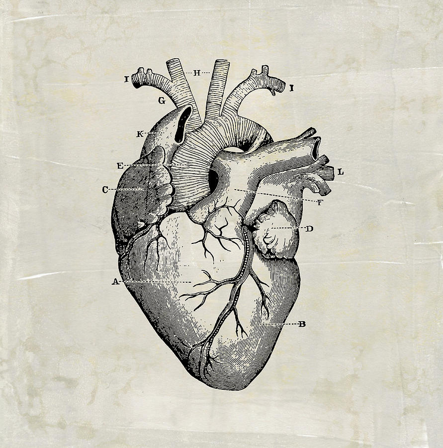
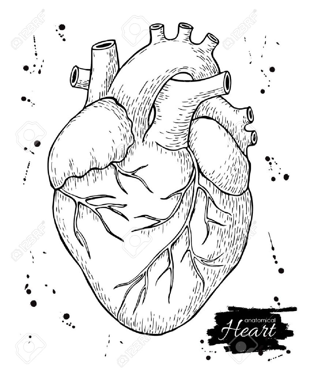
A drawing of the anatomy of the opened normal heart, with english labels. Base (posterior), diaphragmatic (inferior), sternocostal (anterior), and left and right pulmonary surfaces. Web create a curved shape similar to an acorn or apple’s bottom half. The user can show or hide the anatomical labels which provide a useful tool to create illustrations perfectly adapted for teaching. The heart has five surfaces: The right margin is the small section of the right atrium that extends between the superior and inferior vena cava. We will then proceed to shape the heart, slowly refining it. January 29, 2024 | published on: This line is called a guide line. You will erase it later, but in the meantime, it will help you draw a perfect heart.
Web to draw the internal structure of the heart, start by sketching the 2 pulmonary veins to the lower left of the aorta and the bottom of the inferior vena cava slightly to the right of that. You will erase it later, but in the meantime, it will help you draw a perfect heart. Drawing internal anatomy of the heart. Base (posterior), diaphragmatic (inferior), sternocostal (anterior), and left and right pulmonary surfaces. By following the simple steps, you too can easily draw a perfect human heart. The heart has five surfaces: Angle the slightly tampered end of the shape to the left about 120 degrees. Begin the easy heart outline by drawing a straight, vertical line. A drawing of the anatomy of the opened normal heart, with english labels. Web heart pictures, diagram & anatomy | body maps.
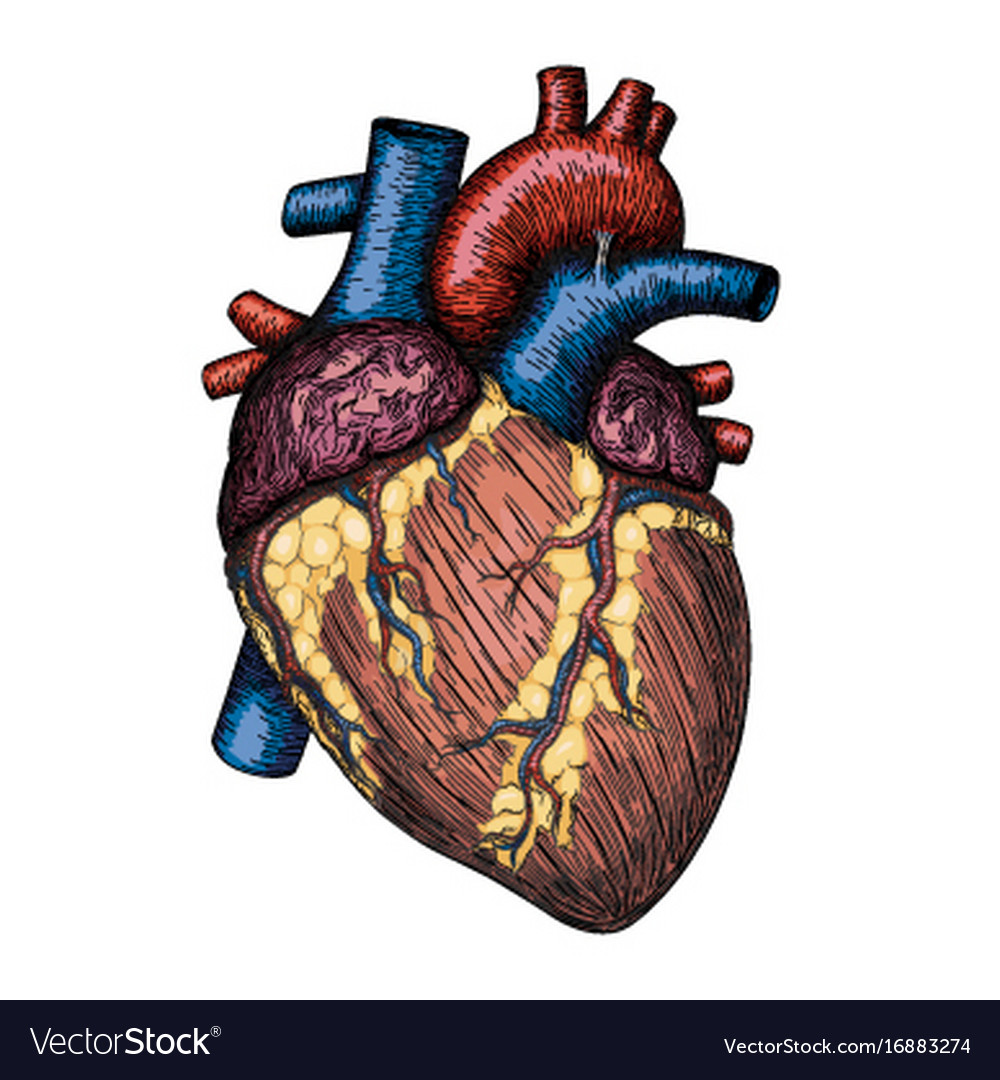
Human heart hand drawn anatomical sketch Vector Image
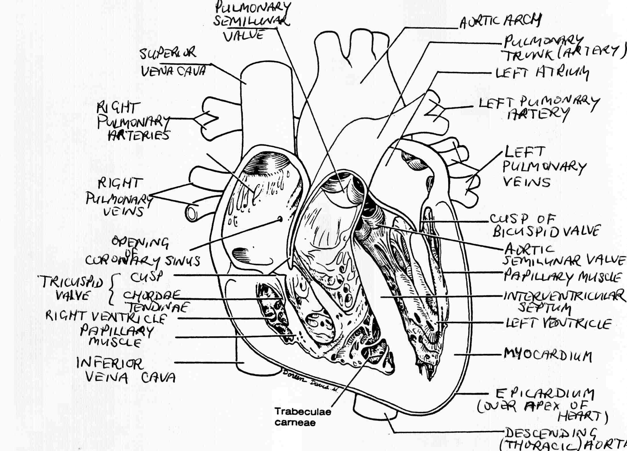
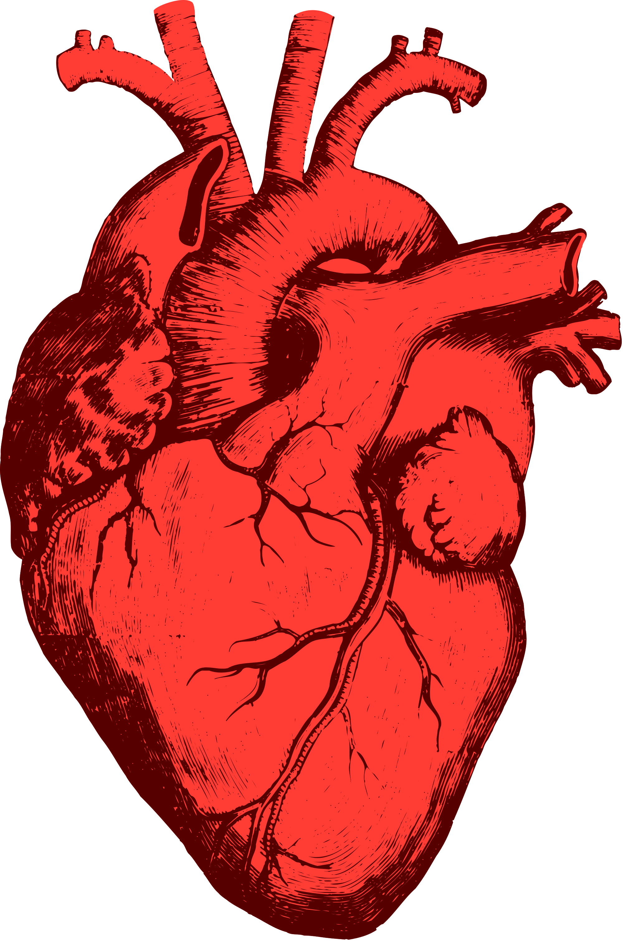
Begin the easy heart outline by drawing a straight, vertical line. We will then proceed to shape the heart, slowly refining it. Drawings of the surface anatomy of the normal heart, anterior and posterior, with english labels. The user can show or hide the anatomical labels which provide a useful tool to create illustrations perfectly adapted for teaching. Web this.
Anatomical Heart Drawing at Explore collection of
Drawing internal anatomy of the heart. [1] the main shape will be the basis for the left and right ventricles. Web this interactive atlas of human heart anatomy is based on medical illustrations and cadaver photography. Drawings of the surface anatomy of the normal heart, anterior and posterior, with english labels. Begin the easy heart outline by drawing a straight,.

How to Draw the Internal Structure of the Heart (with Pictures)
Web create a curved shape similar to an acorn or apple’s bottom half. [1] the main shape will be the basis for the left and right ventricles. Right, left, superior, and inferior: The heart has five surfaces: Drawing internal anatomy of the heart.

anatomic heart (With images) Anatomy art, Medical drawings, Heart diagram
From the openstax anatomy and physiology. Then, fill in the base of the heart with the. Drawing internal anatomy of the heart. From the openstax anatomy and physiology book. You can use a ruler or straight edge.
The best free Structure drawing images. Download from 877 free drawings
Drawing internal anatomy of the heart. By following the simple steps, you too can easily draw a perfect human heart. We will then proceed to shape the heart, slowly refining it. The heart is a mostly hollow, muscular organ composed of cardiac muscles and connective tissue that. Base (posterior), diaphragmatic (inferior), sternocostal (anterior), and left and right pulmonary surfaces.
8 Anatomical Heart Drawings! The Graphics Fairy
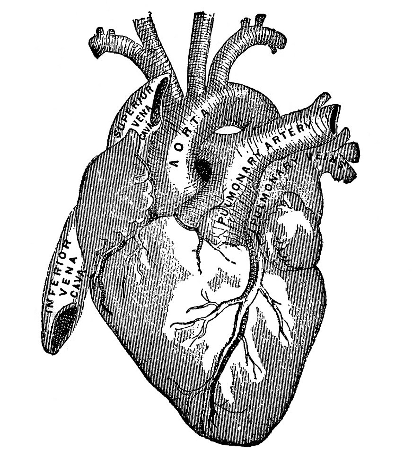
You can use a ruler or straight edge. A drawing of the anatomy of the opened normal heart, with english labels. From the openstax anatomy and physiology. The right margin is the small section of the right atrium that extends between the superior and inferior vena cava. By following the simple steps, you too can easily draw a perfect human.

anatomy medical Google Search in 2020 Medical illustration, Heart
From the openstax anatomy and physiology. Drawing internal anatomy of the heart. Base (posterior), diaphragmatic (inferior), sternocostal (anterior), and left and right pulmonary surfaces. It also has several margins: Begin the easy heart outline by drawing a straight, vertical line.

How to Draw the Internal Structure of the Heart 13 Steps
Web to draw the internal structure of the heart, start by sketching the 2 pulmonary veins to the lower left of the aorta and the bottom of the inferior vena cava slightly to the right of that. From the openstax anatomy and physiology. You will erase it later, but in the meantime, it will help you draw a perfect heart..
Anatomical Drawing Heart at GetDrawings Free download
From the openstax anatomy and physiology book. You can use a ruler or straight edge. We will then proceed to shape the heart, slowly refining it. Web drawings surface anatomy of the heart. Web in this lecture, dr mike shows the two best ways to draw and label the heart!
Human Heart Drawing Free download on ClipArtMag
This line is called a guide line. From the openstax anatomy and physiology book. We will then proceed to shape the heart, slowly refining it. Begin the easy heart outline by drawing a straight, vertical line. Web drawings surface anatomy of the heart.
Begin The Easy Heart Outline By Drawing A Straight, Vertical Line.
By following the simple steps, you too can easily draw a perfect human heart. Web to draw the internal structure of the heart, start by sketching the 2 pulmonary veins to the lower left of the aorta and the bottom of the inferior vena cava slightly to the right of that. January 29, 2024 | published on: It also has several margins:
Drawing Internal Anatomy Of The Heart.
The right margin is the small section of the right atrium that extends between the superior and inferior vena cava. Base (posterior), diaphragmatic (inferior), sternocostal (anterior), and left and right pulmonary surfaces. The heart has five surfaces: From the openstax anatomy and physiology book.
A Drawing Of The Anatomy Of The Opened Normal Heart, With English Labels.
We will then proceed to shape the heart, slowly refining it. The user can show or hide the anatomical labels which provide a useful tool to create illustrations perfectly adapted for teaching. Then, fill in the base of the heart with the. Web this interactive atlas of human heart anatomy is based on medical illustrations and cadaver photography.
Web Create A Curved Shape Similar To An Acorn Or Apple’s Bottom Half.
You will erase it later, but in the meantime, it will help you draw a perfect heart. This line is called a guide line. Web in this lecture, dr mike shows the two best ways to draw and label the heart! Web heart pictures, diagram & anatomy | body maps.