Animal Cell Draw And Label
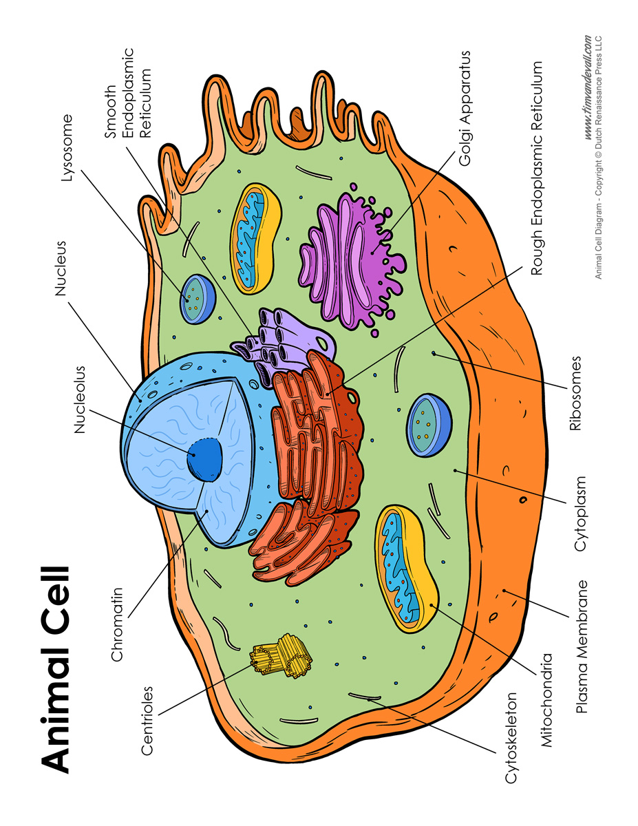
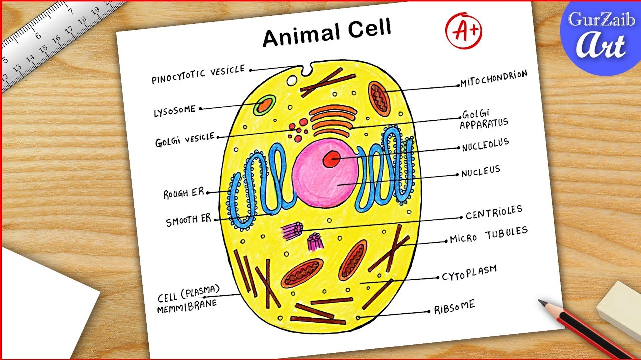
Animal Cell Draw And Label - Next, draw the nucleus by adding a circle inside the membrane with a smaller circle inside it. Name the numbered parts of the cell. The columns to the left and right of the labels contain links to discussions of the particular structures. Download a free printable outline of this video and. Students can print images to help them learn the cell. The next is a black and white version of the first. This schematic represents an idealized animal cell, e.g., a liver cell. Web use this convenient study aid in preparation for your upcoming test or quiz. Web how to draw a animal cell easy and step by step. Animal cells are the basic unit of life in organisms of the kingdom animalia.
They are eukaryotic cells, meaning that they have a true nucleus and specialized structures called organelles that carry out different functions. Diagram of a plant cell. How proteins are packaged for transport. Name the numbered parts of the cell. Unlike the animal cell lacking the cell wall, plant cells have a. The animal kingdom contains the largest number of species on the entire earth. Students can print images to help them learn the cell. The next is a black and white version of the first. Observe and identify differences between cells and cell structures under low and high magnification and record your observations. Where, prokaryotes are just bacteria and archaea, eukaryotes are literally everything else.
Review your understanding of cell parts and functions in this free article aligned to ngss standards. These printables a free for subscribing members of tim’s printables. Name the numbered parts of the cell. Students can print images to help them learn the cell. All organisms are made up of cells (or in some cases, a single cell). This schematic represents an idealized animal cell, e.g., a liver cell. The animal kingdom contains the largest number of species on the entire earth. 56k views 6 years ago. After completing the lab, the student will be able to: All cells have a cell membrane that separates the inside and the outside of the cell, and controls what goes in and comes out.

Animal Cell Diagrams Labeled Printable 101 Diagrams
These printables a free for subscribing members of tim’s printables. Most cells are very small; The animal cell function includes energy production, transportation, protein synthesis, cellular communication, movement, and. By following the simple steps, you too can easily draw a perfect animal cell. Kids can learn about the animal cell by labeling & coloring this free handout!

How To Draw Animal Cell Diagram Labeled Functions and Diagram
All cells have a cell membrane that separates the inside and the outside of the cell, and controls what goes in and comes out. After completing the lab, the student will be able to: These printables a free for subscribing members of tim’s printables. Its outer coating is a semipermeable cell membrane. This entry was posted on may 9, 2023.

Animal Cell Diagram drawing How To Draw Animal Cell Labeled Science
Download a free printable outline of this video and. Below is the answer key: Web the animal cell diagram is widely asked in class 10 and 12 examinations and is beneficial to understand the structure and functions of an animal. Animal cells are the basic unit of life in organisms of the kingdom animalia. Students can print images to help.
Animal Cell Diagram Labeled Tim van de Vall
Which organelle contains its own dna? This worksheet helps students learn the parts of the cell. Where, prokaryotes are just bacteria and archaea, eukaryotes are literally everything else. Web label the parts of the plant and animal cell. The animal cell function includes energy production, transportation, protein synthesis, cellular communication, movement, and.

How to Draw an Animal Cell Really Easy Drawing Tutorial
Below is the answer key: Web how to draw a animal cell easy and step by step. Next, draw the nucleus by adding a circle inside the membrane with a smaller circle inside it. Web a compilation of plant and animal cell images with organelles and major structures labeled. Which organelle contains its own dna?
Animal Cell Structure Carlson Stock Art
Download a free printable outline of this video and. By following the simple steps, you too can easily draw a perfect animal cell. Where, prokaryotes are just bacteria and archaea, eukaryotes are literally everything else. This worksheet helps students learn the parts of the cell. They are eukaryotic cells, meaning that they have a true nucleus and specialized structures called.
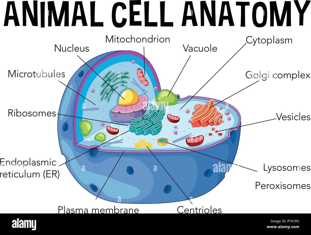
Diagram of animal cell anatomy illustration Stock Vector Image & Art
After completing the lab, the student will be able to: How proteins are packaged for transport. All organisms are made up of cells (or in some cases, a single cell). This entry was posted on may 9, 2023 by anne helmenstine (updated on january 29, 2024) an animal cell lacks a cell wall or chloroplasts. Web label the parts of.

What Is An Animal Cell? Facts, Pictures & Info For Kids & Students.
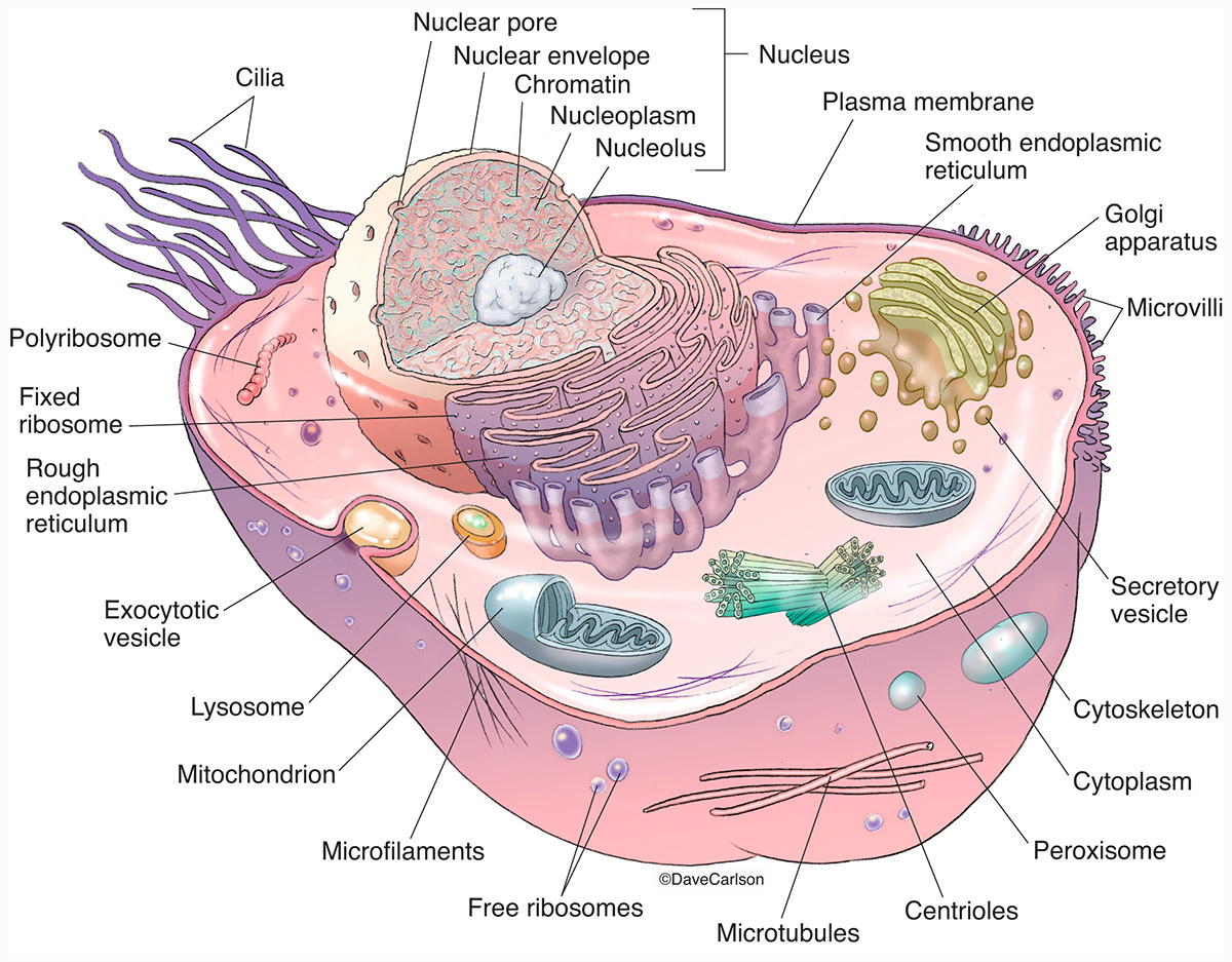
Web a compilation of plant and animal cell images with organelles and major structures labeled. Web the most important structures of plant and animal cells are shown in the diagrams below, which provide a clear illustration of how much these cells have in common. These printables a free for subscribing members of tim’s printables. An animal cell diagram is useful.
Animal Cell Diagram Drawing CBSE easy way labeled Science project
Animal cells are the fundamental units of life in protozoa and multicellular animals. How proteins are packaged for transport. Web a compilation of plant and animal cell images with organelles and major structures labeled. Name the numbered parts of the cell. Animal cells are the basic unit of life in organisms of the kingdom animalia.
Animal Cell Drawing at GetDrawings Free download
The cell organelles are enclosed by the plasma membrane including the cell nucleus. Animal cells are the fundamental units of life in protozoa and multicellular animals. Web how to draw a animal cell easy and step by step. Web label the parts of the plant and animal cell. Download a free printable outline of this video and.
The Animal Kingdom Contains The Largest Number Of Species On The Entire Earth.
Web how to draw a animal cell easy and step by step. How proteins are packaged for transport. An animal cell diagram is useful for understanding the structure and functioning of an animal. Web an animal cell is a eukaryotic cell that lacks a cell wall, and it is enclosed by the plasma membrane.
All Cells Have A Cell Membrane That Separates The Inside And The Outside Of The Cell, And Controls What Goes In And Comes Out.
Get free printable coloring page of this drawing. Web the animal cell diagram is widely asked in class 10 and 12 examinations and is beneficial to understand the structure and functions of an animal. Web label the parts of the plant and animal cell. Observe and identify differences between cells and cell structures under low and high magnification and record your observations.
Eukaryotic Cells Are Larger, More Complex, And Have Evolved More Recently Than Prokaryotes.
Where, prokaryotes are just bacteria and archaea, eukaryotes are literally everything else. The cell is the basic unit of life. This entry was posted on may 9, 2023 by anne helmenstine (updated on january 29, 2024) an animal cell lacks a cell wall or chloroplasts. Animal cells are the fundamental units of life in protozoa and multicellular animals.
This Page Is A Draft And Is Under Active Development.
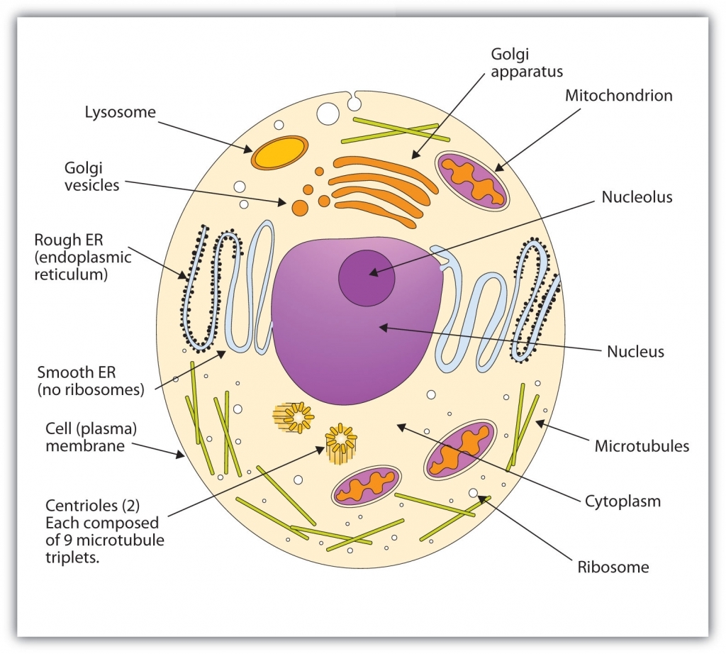
Below is the answer key: This schematic represents an idealized animal cell, e.g., a liver cell. Review your understanding of cell parts and functions in this free article aligned to ngss standards. Students also label a diagram showing how proteins are produced by ribosomes, transported via the endoplasmic reticulum, and finally packaged by the golgi apparatus.