Animal Cell Drawing And Label
Animal Cell Drawing And Label - This schematic represents an idealized animal cell, e.g., a liver cell. Finally, an unlabeled version of the diagram is included at the bottom of the page, in color and black and white. Web one can observe the golgi apparatus in the labeled animal cell parts diagram. Web the smallest cell in the animal is a neuron that is just 100 micrometers in diameter. A plasma membrane encloses the cell contents of both plant and animal cells, but it is the outer coating of an animal cell. An animal cell is a eukaryotic cell that lacks a cell wall, and it is enclosed by the plasma membrane. Dna contains all the instructions for making proteins, which control all of the body’s activities. The cell membrane of an animal cell is not a perfect circle. This is a barrier that surrounds the cell and holds it together. Web diagram of animal cell.
Web overview of animal and plant cells (opens a modal) practice. The third and fourth diagrams are animal cell diagram worksheets. Forms the external barrier of our body that provides protection. The main function of this golgi complex is to receive the proteins synthesized in the er and transform it into more complex proteins. They are different from plant cells in that they do contain cell walls and chloroplast. Web the animal cell that forms the epidermis is referred to as “skin cells.”. These are the langerhans cells, merkel cells, keratinocytes, and melanocytes. You can make the circle misshapen or oblong. All organisms are made up of cells (or in some cases, a single cell). These h5p resources are made available openly with the cc by license.
The various cell organelles present in an animal cell are clearly marked in the animal cell diagram provided below. Structure, parts, functions, labeled diagram. [1] also know that the membrane is not a rigid cell wall like in plant cells. It performs the specific functions like protection, absorption, and secretion. These are the langerhans cells, merkel cells, keratinocytes, and melanocytes. Web labeled diagram of a typical animal cell nucleus. Explore the various organelles and their roles in maintaining homeostasis. The columns to the left and right of the labels contain links to discussions of the particular structures. The cells can be squamous, columnar, or cuboidal in shape. The animal cell function includes energy production, transportation, protein synthesis, cellular communication, movement, and maintenance of structure.

Animal Cell Drawing at GetDrawings Free download
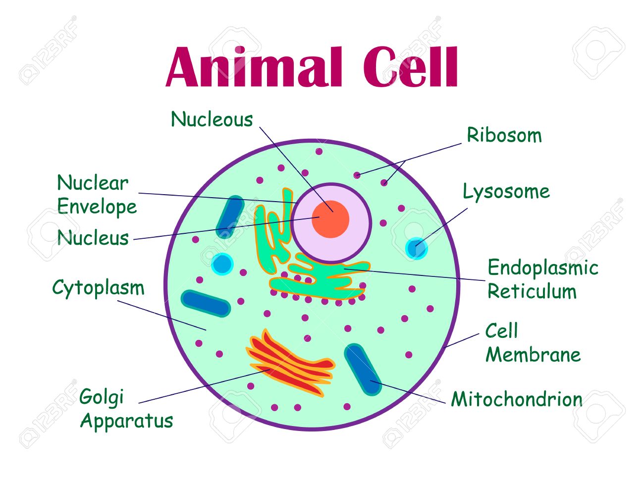
The columns to the left and right of the labels contain links to discussions of the particular structures. Web diagram of animal cell. The outermost part of the cell, which is shown as an outline of the cell, is labeled cell membrane. The important part is that it does not have any sharp edges. Unlike plant cells, animal cells do.

How to Draw an Animal Cell Really Easy Drawing Tutorial
These cells are further described as being eukaryotic cells. Finally, an unlabeled version of the diagram is included at the bottom of the page, in color and black and white. Web the diagram given below depicts the structural organization of the animal cell. This genetic information is called deoxyribonucleic acid (dna). The cell membrane of an animal cell is not.

Animal Cell Definition Structure Parts Functions Labeled Diagram Riset
All organisms are made up of cells (or in some cases, a single cell). This genetic information is called deoxyribonucleic acid (dna). Due to the absence of cell walls, animal cells are less rigid and can take up various shapes. The animal cell diagram is widely asked in class 10 and 12 examinations and is beneficial to understand the structure.

What is a cell? Facts
The third and fourth diagrams are animal cell diagram worksheets. Web on the left is a circle representing an animal cell. Web an animal cell diagram is useful for understanding the structure and functioning of an animal. They are different from plant cells in that they do contain cell walls and chloroplast. All organisms are made up of cells (or.

How To Draw Animal Cell Diagram Labeled Functions and Diagram
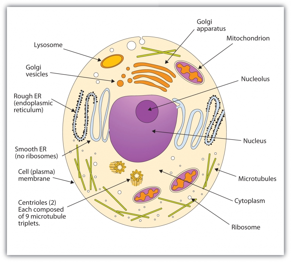
The main function of this golgi complex is to receive the proteins synthesized in the er and transform it into more complex proteins. Structure, parts, functions, labeled diagram. Most cells are very small; Animal cell diagram detailing the various organelles. Quiz yourself by filling in the blanks.

What Is An Animal Cell? Facts, Pictures & Info For Kids & Students.
This genetic information is called deoxyribonucleic acid (dna). The cell is the basic unit of life. The various cell organelles present in an animal cell are clearly marked in the animal cell diagram provided below. Web the diagram given below depicts the structural organization of the animal cell. The cell organelles are enclosed by the plasma membrane including the cell.
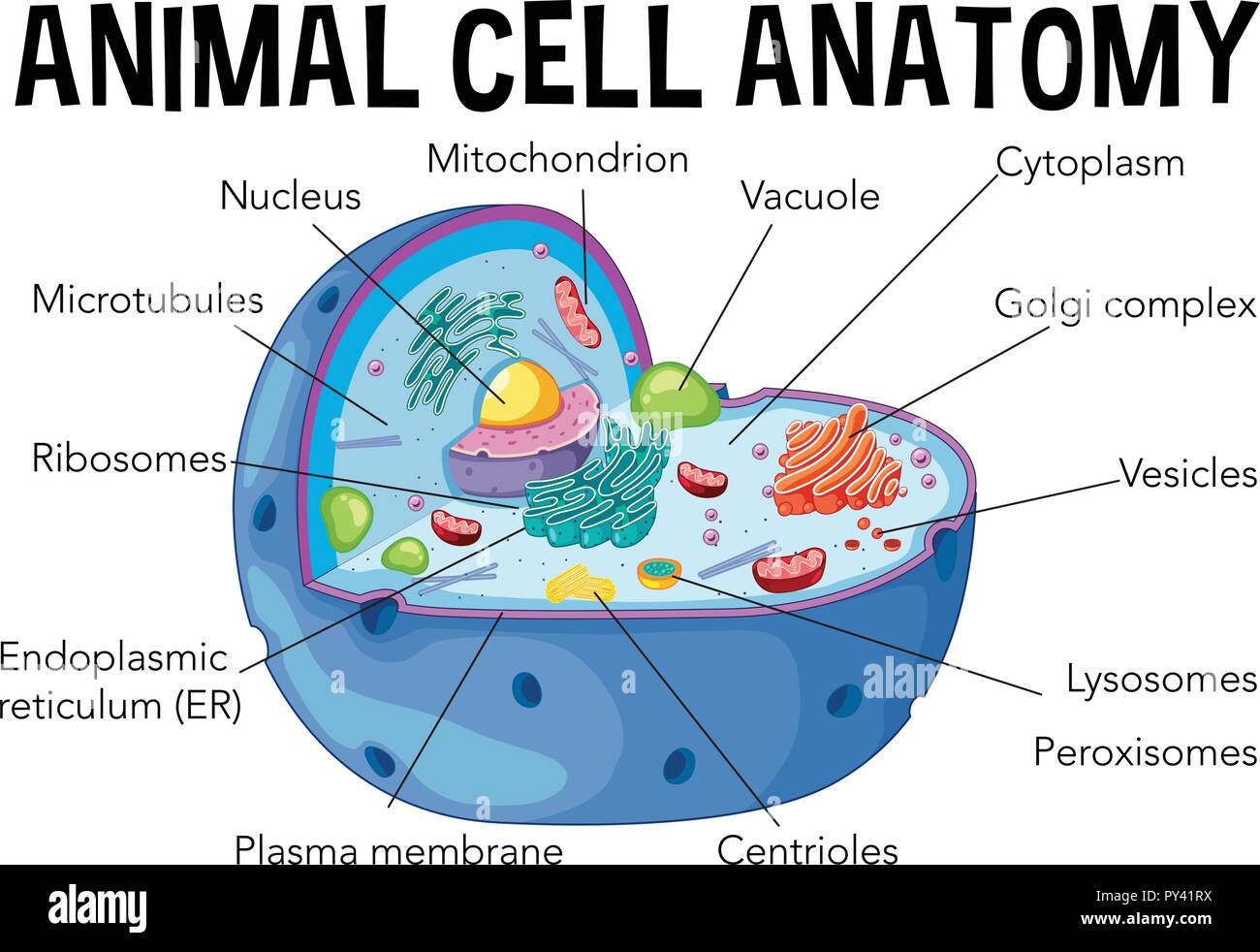
Diagram of animal cell anatomy illustration Stock Vector Image & Art
They are different from plant cells in that they do contain cell walls and chloroplast. Extracellular structures and intercellular junctions get 3 of 4 questions to level up! By go life science posted on december 20, 2022 october 17, 2023. [1] also know that the membrane is not a rigid cell wall like in plant cells. You can make the.
Animal Cells Drawing at GetDrawings Free download
Web overview of animal and plant cells (opens a modal) practice. This schematic represents an idealized animal cell, e.g., a liver cell. Structure, parts, functions, labeled diagram. Various animal cell parts known as animal cell organelles have specialized. Web the diagram given below depicts the structural organization of the animal cell.
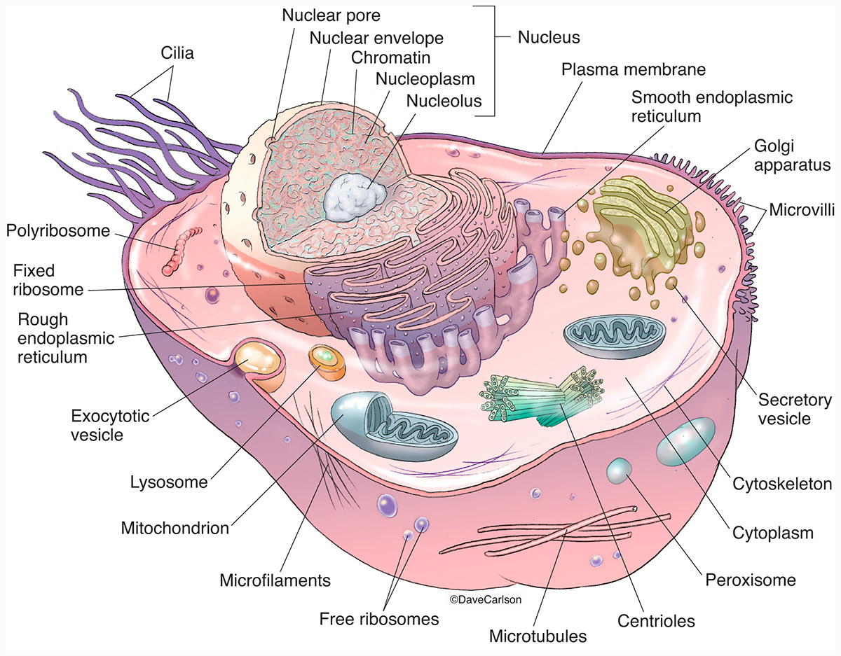
Animal Cell Structure Carlson Stock Art
Due to the absence of cell walls, animal cells are less rigid and can take up various shapes. The animal cell diagram is widely asked in class 10 and 12 examinations and is beneficial to understand the structure and functions of an animal. Web diagram of animal cell. Forms the external barrier of our body that provides protection. These h5p.
Animal Cell Drawing at GetDrawings Free download
These are the langerhans cells, merkel cells, keratinocytes, and melanocytes. A plasma membrane encloses the cell contents of both plant and animal cells, but it is the outer coating of an animal cell. Due to the absence of cell walls, animal cells are less rigid and can take up various shapes. The cell is the basic unit of life. Finally,.
Level Up On The Above Skills And Collect Up To 160 Mastery Points Start Quiz.
Web animal cells are the building blocks that make up all living organisms in the kingdom animalia. The cell organelles are enclosed by the plasma membrane including the cell nucleus. Web animal cell structure. The animal cell diagram is widely asked in class 10 and 12 examinations and is beneficial to understand the structure and functions of an animal.
Unlike The Eukaryotic Cells Of Plants And Fungi, Animal Cells Do Not Have A Cell Wall.
Explore the various organelles and their roles in maintaining homeostasis. Animal cells have centrosomes (or a pair of centrioles), and lysosomes, whereas plant cells do not. Blank animal cell diagram worksheet. These h5p resources are made available openly with the cc by license.
Web Overview Of Animal And Plant Cells (Opens A Modal) Practice.
The third and fourth diagrams are animal cell diagram worksheets. These are the langerhans cells, merkel cells, keratinocytes, and melanocytes. An animal cell is a eukaryotic cell that lacks a cell wall, and it is enclosed by the plasma membrane. The cells can be squamous, columnar, or cuboidal in shape.
Web The Smallest Cell In The Animal Is A Neuron That Is Just 100 Micrometers In Diameter.
Web learn about the structure and function of animal cells, the basic unit of life in animals. Finally, an unlabeled version of the diagram is included at the bottom of the page, in color and black and white. Extracellular structures and intercellular junctions get 3 of 4 questions to level up! Likewise, the largest cell is the egg of an ostrich that can be up to 150 millimeters in diameter and weigh about 3 pounds.