Brain And Spinal Cord Drawing
Brain And Spinal Cord Drawing - The central nervous system (the brain and spinal cord) and the peripheral nervous system (the nerves outside the brain and spinal cord). The other main division is the peripheral nervous system (pns). What is the brain made of? Web it will be costly — cross estimates they need to raise at least $2 million — but the potential upside could be priceless. Web some sensory messages are immediately acted on by the spinal cord, without any input from the brain. Web brain and spinal cord drawing stock photos and images. (869) see brain and spinal cord drawing stock video clips. Withdrawal from a hot object and the knee jerk are two examples. Affected taste in the anterior 2/3 of the tongue. The brain is one of the most fun parts of the body to draw.
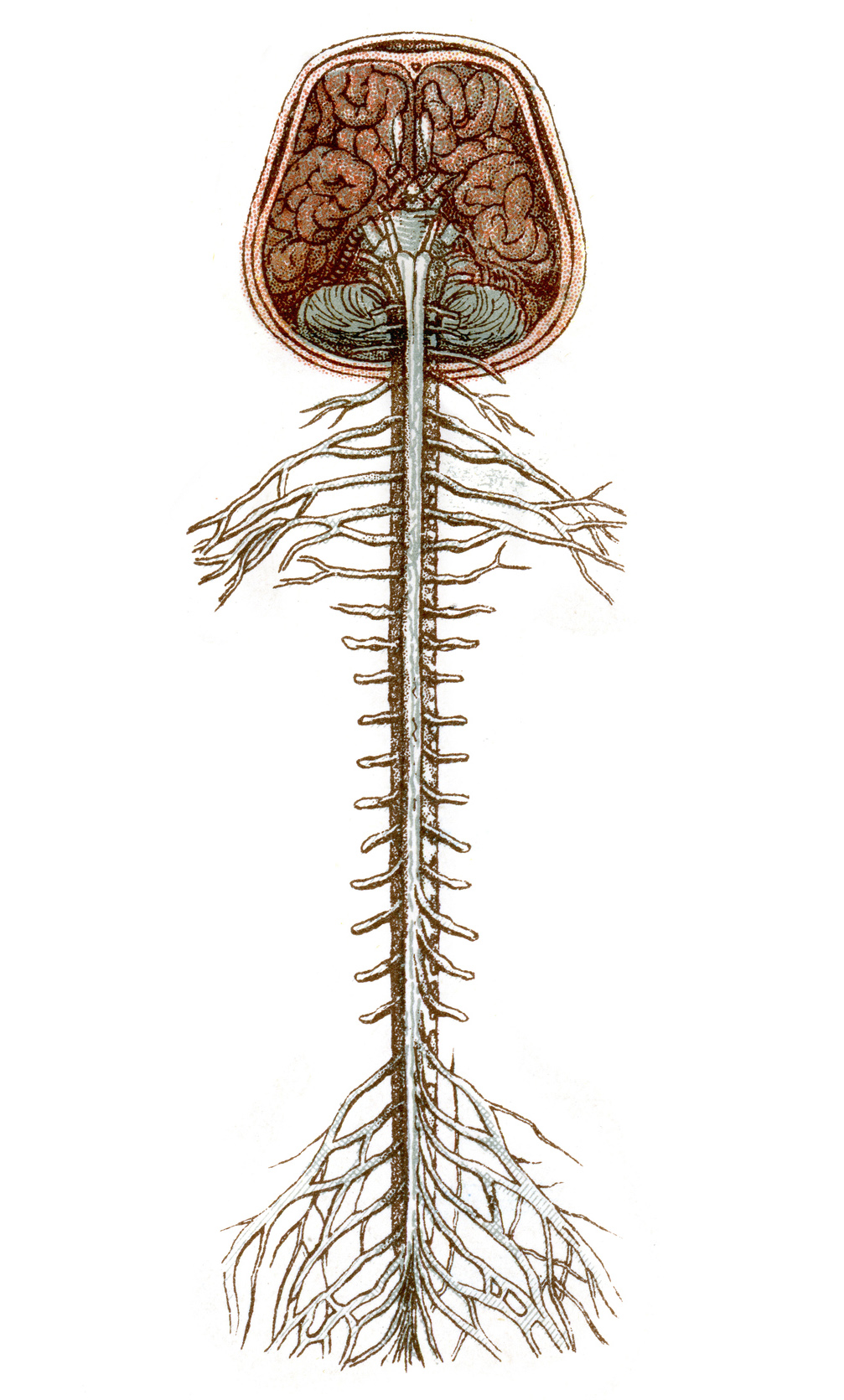
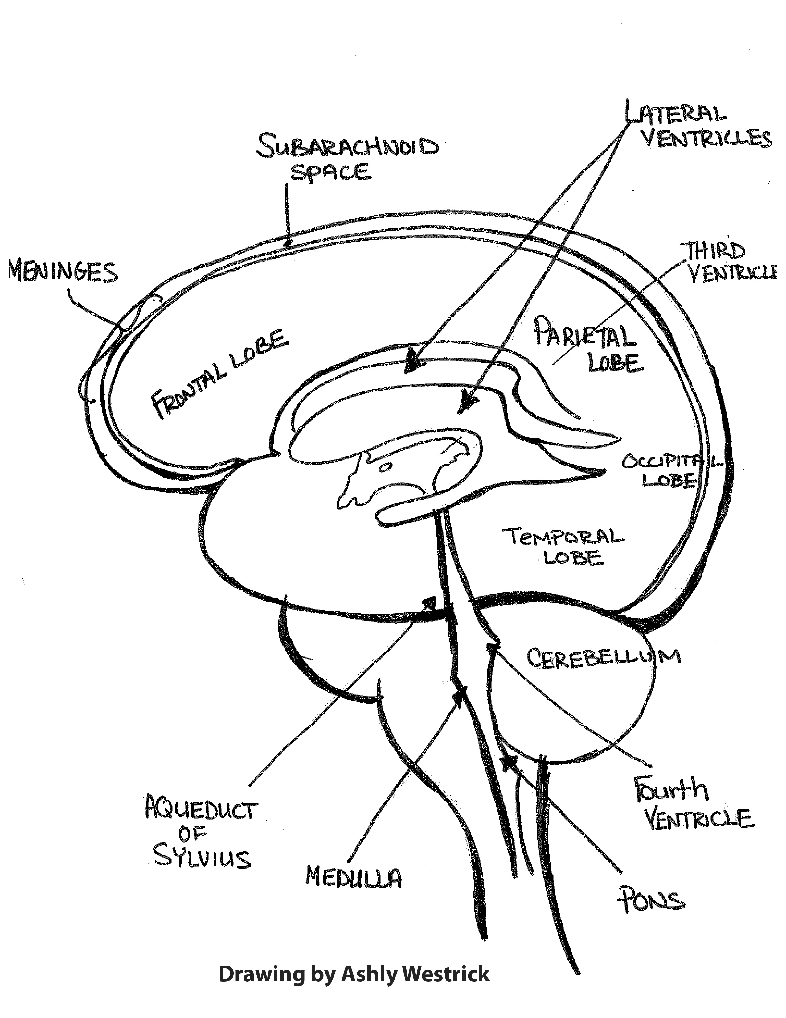
The brain itself is a not a muscle. Both of these are protected by three layers of membranes known as meninges. If you want more of a challenge, include anatomical parts, such as the brain stem and cerebellum. The brain is one of the most fun parts of the body to draw. Web together, the brain and spinal cord that extends from it make up the central nervous system, or cns. The central nervous system (the brain and spinal cord) and the peripheral nervous system (the nerves outside the brain and spinal cord). Web schematic drawing of subcortical diencephalic and mesencephalic structures. What is the brain made of? Color vector illustration of children's activity coloring book pages with pictures of orange spine. It extends from the foramen magnum at the base of the skull to the l1/l2 vertebra where it terminates as the conus medullaris (medullary cone).
Describe the types of techniques available to clinicians and researchers to image or scan the brain. Anatomy of the vascular system engraving antique illustration, published 1851. Web schematic drawing of subcortical diencephalic and mesencephalic structures. Web the brain is found in the cranial cavity, while the spinal cord is found in the vertebral column. The brain is one of the most fun parts of the body to draw. Web some sensory messages are immediately acted on by the spinal cord, without any input from the brain. You can make it as simple as you like by drawing lots of squiggles and keeping the shape round. The drawing below (figure 8.5.2) shows the central nervous system as one of two main divisions of the total nervous system. The brain is contained within the cranial cavity of the skull, and the spinal cord is contained. Get inspired to create stunning illustrations that capture the intricacies and beauty of this vital part of the human body.

Illustration, human brain, spinal cord Stock Image C005/6988
Both are protected by three layers of meninges ( dura , arachnoid, and pia mater ). When a sensory message meets certain parameters, the spinal cord initiates an automatic reflex. Web some sensory messages are immediately acted on by the spinal cord, without any input from the brain. Weighing about 3 pounds in the average adult, the brain is about.

Human brain and spinal cord, illustration Stock Image F013/2601
When a sensory message meets certain parameters, the spinal cord initiates an automatic reflex. What is the brain made of? Affected taste in the anterior 2/3 of the tongue. April 17, 2024 fact checked. Web the secret in both these cases is a system of flexible electrodes implanted directly on the patients' spinal cords.

Brain And Spinal Cord Diagram Anatomy Chart Of Spinal Cord Labeled
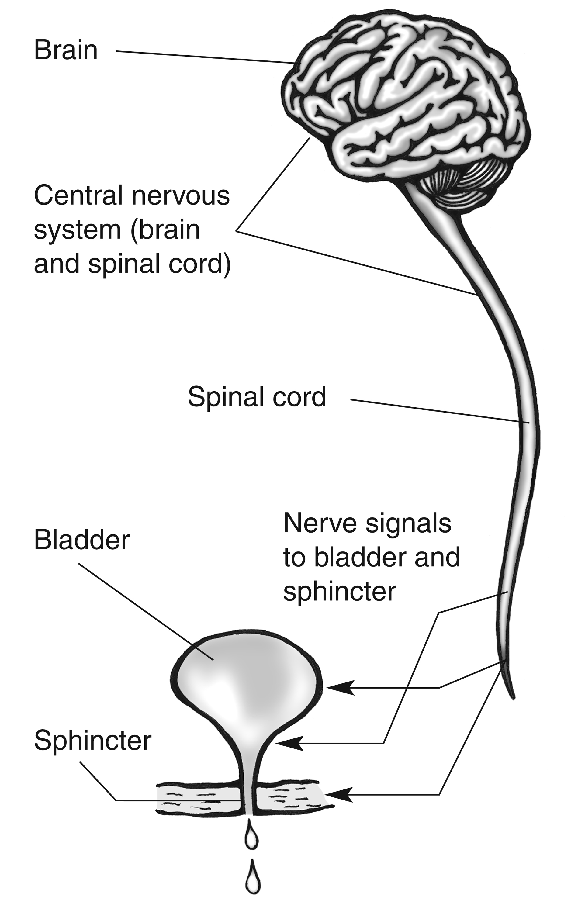
For further protection, the brain is encased within the hard bones of the skull, while the spinal cord is protected with the bony vertebrae of our backbones. Web resources / anatomical drawings / brain and spinal cord. The brain generates commands for target tissues and the spinal cord acts as a conduit, connecting the brain to peripheral tissues via the.
Duke Neurosciences Lab 2 Spinal Cord & Brainstem Surface and
Old chromolithograph illustration of human brain and spinal cord. Web the brain is found in the cranial cavity, while the spinal cord is found in the vertebral column. The metencephalon includes the pons and the cerebellum. “we would treat not just alzheimer’s,” she says, “but also any kind of dementia: Web schematic drawing of subcortical diencephalic and mesencephalic structures.
Brain And Spinal Cord Drawing
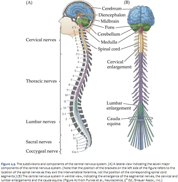
Describe the types of techniques available to clinicians and researchers to image or scan the brain. The drawing below (figure 8.5.2) shows the central nervous system as one of two main divisions of the total nervous system. The cerebrum, the diencephalon, the brain stem, and the cerebellum. Web the brain and the spinal cord are the central nervous system, and.

Human brain and spinal cord, artwork Stock Image F010/1846
Web identify the spinal cord, cerebral lobes, and other brain areas on a diagram of the brain. Identify the hemispheres and lobes of the brain. The cns and pns work together to control virtually. Weighing about 3 pounds in the average adult, the brain is about 60% fat. “we would treat not just alzheimer’s,” she says, “but also any kind.
The spinal cord Queensland Brain Institute University of Queensland
The cerebrum, the diencephalon, the brain stem, and the cerebellum. The spinal cord is a single structure, whereas the adult brain is described in terms of four major regions: Weighing about 3 pounds in the average adult, the brain is about 60% fat. Drawing of the outline of a body showing the nervous system with descriptions of each of the.

Central Nervous System Diagram Brain And Spinal Cord / A Lateral view
Web brain and spinal cord drawing stock photos and images. The brain generates commands for target tissues and the spinal cord acts as a conduit, connecting the brain to peripheral tissues via the pns. Reviewed/revised may 2023 | modified jun 2023. Explore unique and artistic ways to draw the spinal cord. Web some sensory messages are immediately acted on by.
Human Brain Functions, Parts and More Sciencemojo
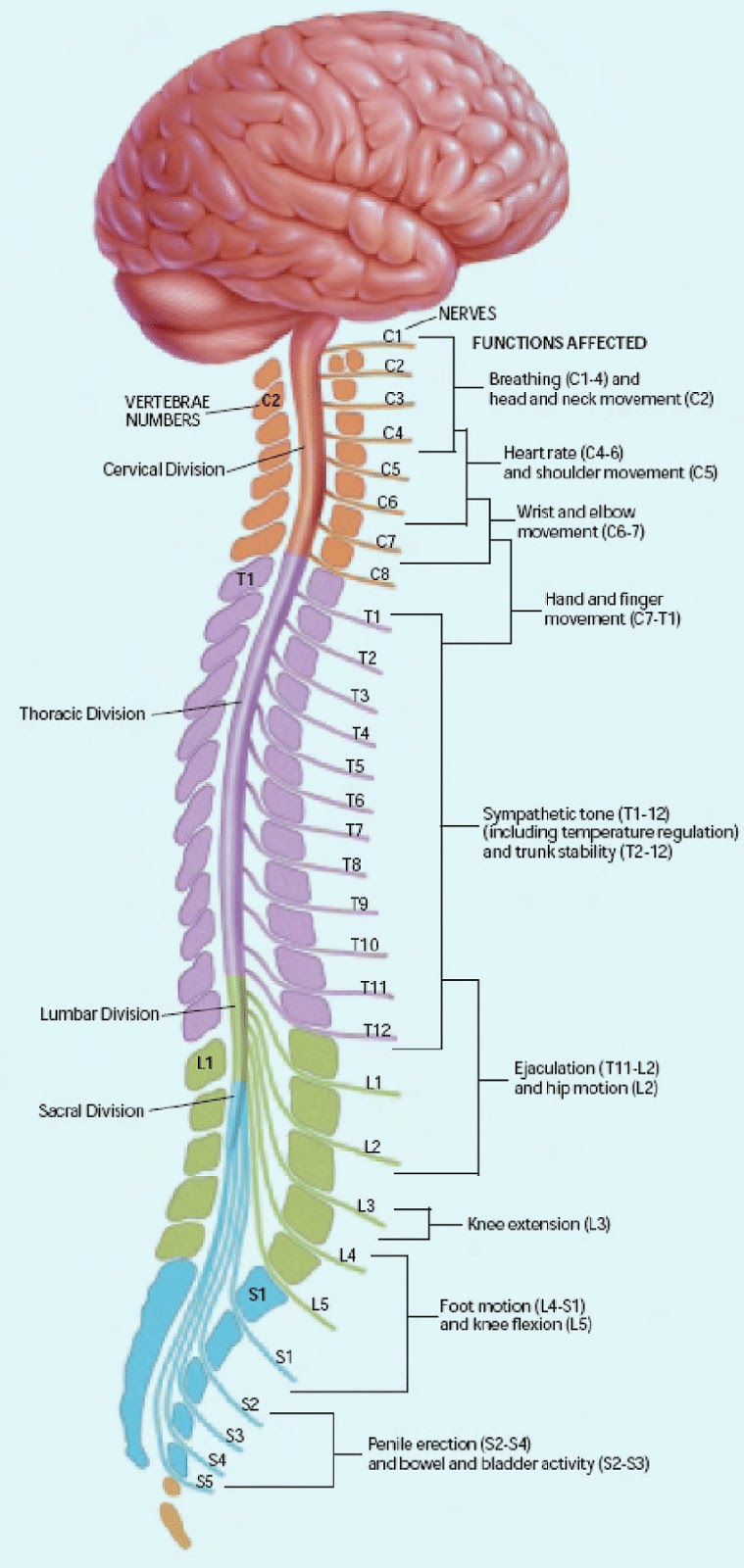
Describe the basic functions of the spinal cord, cerebral lobes, and other brain areas. Explore unique and artistic ways to draw the spinal cord. While the length of the spinal cord varies from one individual to another, it is usually longer in males (approximately 45 cm) than it is in females (approximately 42 cm). Weighing about 3 pounds in the.
Brain 101 An Overview of the Anatomy and Physiology of the Brain
The brain itself is a not a muscle. Identify the hemispheres and lobes of the brain. Web the spinal cord is a cylindrical mass of neural tissue extending from the caudal aspect of the medulla oblongata of the brainstem to the level of the first lumbar vertebra (l1). While the length of the spinal cord varies from one individual to.
Describe The Basic Functions Of The Spinal Cord, Cerebral Lobes, And Other Brain Areas.
The cns and pns work together to control virtually. Web it will be costly — cross estimates they need to raise at least $2 million — but the potential upside could be priceless. Web resources / anatomical drawings / brain and spinal cord. The spinal cord is a single structure, whereas the adult brain is described in terms of four major regions:
The Nervous System Has 2 Distinct Parts:
April 17, 2024 fact checked. Weighing about 3 pounds in the average adult, the brain is about 60% fat. The brain generates commands for target tissues and the spinal cord acts as a conduit, connecting the brain to peripheral tissues via the pns. Both are protected by three layers of meninges ( dura , arachnoid, and pia mater ).
The Brain Is Contained Within The Cranial Cavity Of The Skull, And The Spinal Cord Is Contained.
Cut outs | vectors | black & white. 3d interactive model of the nervous system including: Describe the types of techniques available to clinicians and researchers to image or scan the brain. Withdrawal from a hot object and the knee jerk are two examples.
By The Manual's Editorial Staff.
Reviewed/revised may 2023 | modified jun 2023. You can make it as simple as you like by drawing lots of squiggles and keeping the shape round. The brain itself is a not a muscle. Als, parkinson’s, multiple sclerosis, spinal cord injury, any kind of condition where nerve cells are dying.”.