Centrioles Drawing Easy
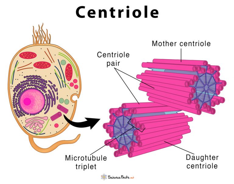
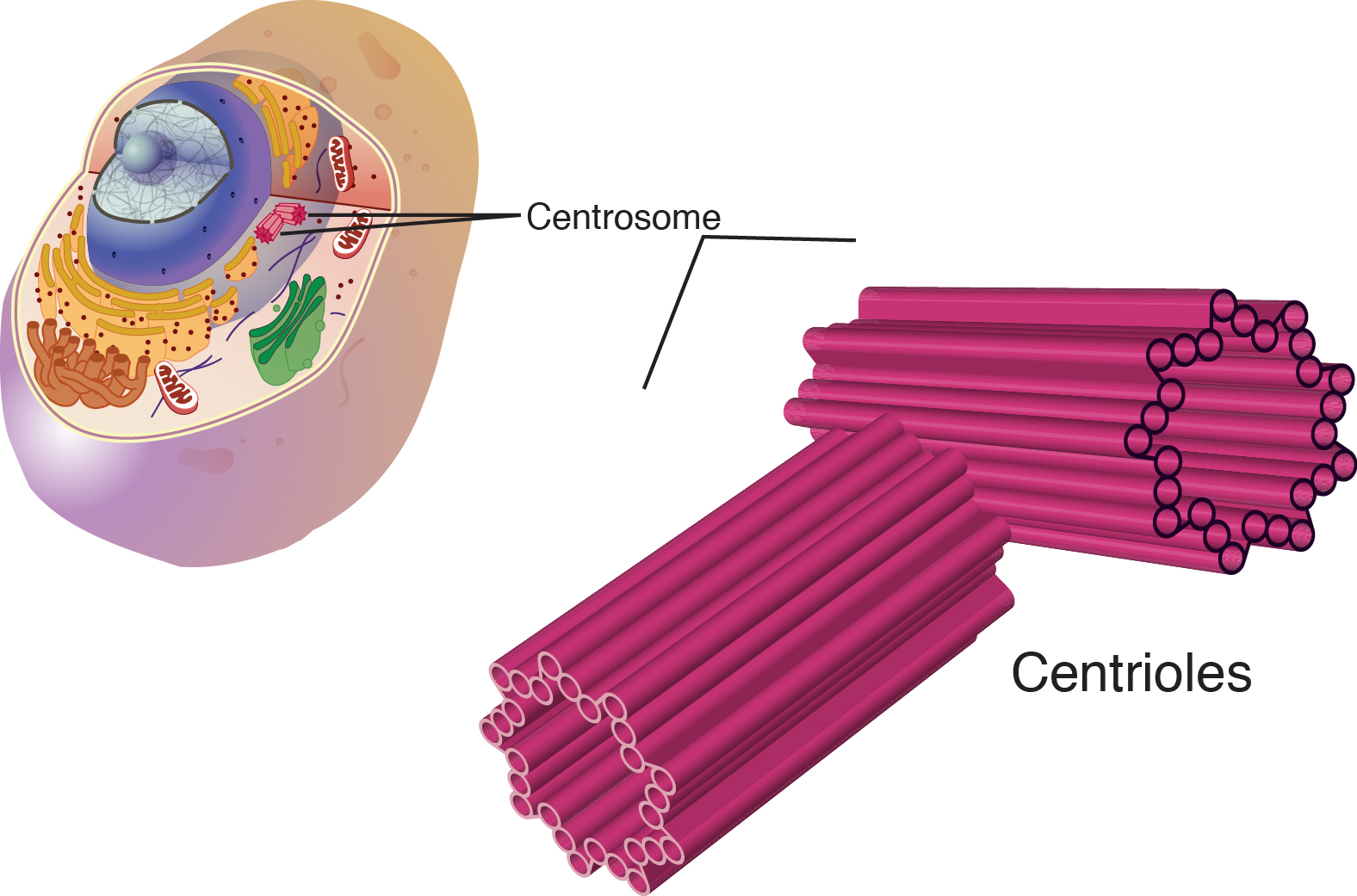
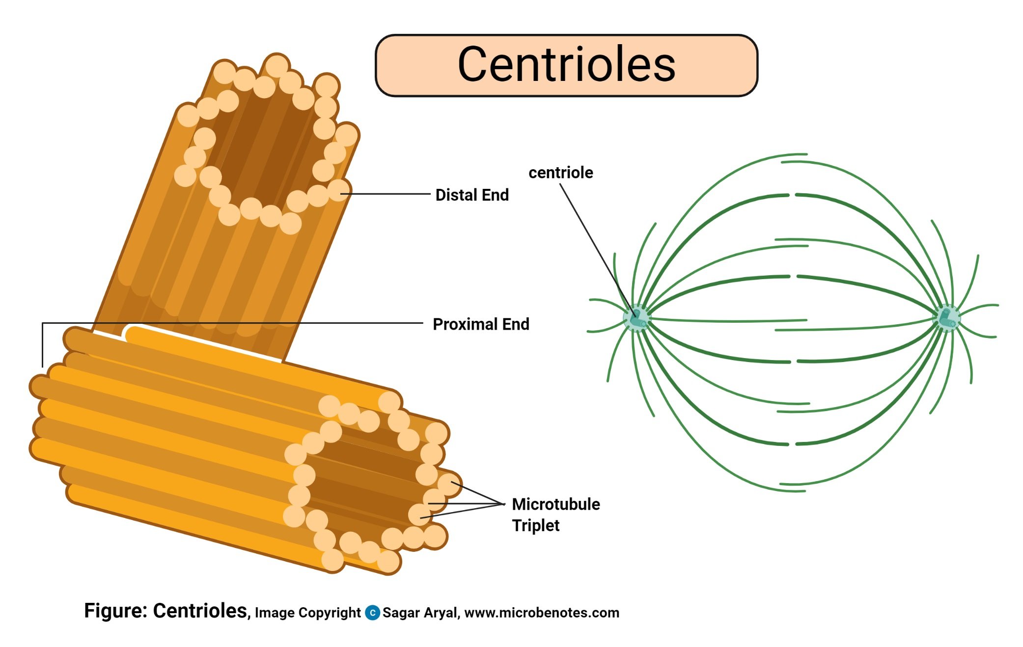
Centrioles Drawing Easy - How to draw centriole | drawing structure of centroiole step by step in easy wayhello friends in this video i tell you about how to draw centrioles. A centriole is a small structure made of microtubules which exists as part of the centrosome, which helps organize microtubules in the body. How to draw structure of centriole | how to draw centrioleshello friends in this video i tell you about how to draw centrioles. It consists of two centrioles — oriented at right angles to each other — embedded in a mass of amorphous material containing more than 100 different proteins.it is duplicated during s phase of the cell cycle. The cell membrane of an animal cell is not a perfect circle. Just before mitosis, the two centrosomes. Web structure of centrioles. When discussing analogies, it often involves identifying shared characteristics or functions. They are visible under a light microscope, but the details of the centriole structure were revealed only under an. Typically, a eukaryotic cell has one centriole that is at a right angle to a second centriole in the.
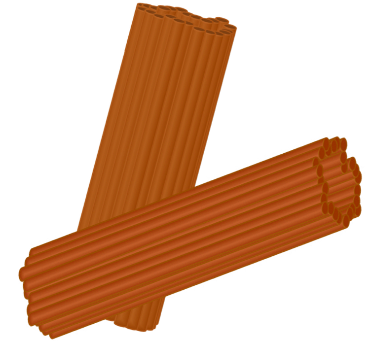
The important part is that it does not have any sharp edges. Web a centriole is a cylindrical cell structure involved in cell division, organizing microtubules for chromosome segregation. The cell membrane of an animal cell is not a perfect circle. You can make the circle misshapen or oblong. Centrioles play a crucial role in. When discussing analogies, it often involves identifying shared characteristics or functions. Web size and shape. A pair of centrioles lies in a common specialized part of the. Centrioles are visible under a light microscope but can be viewed in detail only under an electron microscope. They are visible under a light microscope, but the details of the centriole structure were revealed only under an.
Centrioles are visible under a light microscope but can be viewed in detail only under an electron microscope. They are visible under a light microscope, but the details of the centriole structure were revealed only under an. A centriole is the main unit that creates and anchors microtubules in the cell. Web size and shape. The important part is that it does not have any sharp edges. How to draw centriole | drawing structure of centroiole step by step in easy wayhello friends in this video i tell you about how to draw centrioles. A pair of centrioles lies in a common specialized part of the. Centrioles play a crucial role in. Web a centriole is a cylindrical cell structure involved in cell division, organizing microtubules for chromosome segregation. Just before mitosis, the two centrosomes.
Centriole Definition, Structure, & Functions, with Diagram
The important part is that it does not have any sharp edges. Centrioles play a crucial role in. Centrioles are visible under a light microscope but can be viewed in detail only under an electron microscope. Web structure of centrioles. Web a centriole is a cylindrical cell structure involved in cell division, organizing microtubules for chromosome segregation.
Cell Division II Biology Visionlearning
How to draw structure of centriole | how to draw centrioleshello friends in this video i tell you about how to draw centrioles. When discussing analogies, it often involves identifying shared characteristics or functions. It consists of two centrioles — oriented at right angles to each other — embedded in a mass of amorphous material containing more than 100 different.
Centrioles Drawing
[1] also know that the membrane is not a rigid cell wall like in plant cells. Centrioles play a crucial role in. You can make the circle misshapen or oblong. The centrosome is located in the cytoplasm usually close to the nucleus. Web size and shape.
EduPic Cell Drawings
When discussing analogies, it often involves identifying shared characteristics or functions. The important part is that it does not have any sharp edges. It consists of two centrioles — oriented at right angles to each other — embedded in a mass of amorphous material containing more than 100 different proteins.it is duplicated during s phase of the cell cycle. Just.
Centrioles Easy To Draw , Free Transparent Clipart ClipartKey
[1] also know that the membrane is not a rigid cell wall like in plant cells. It consists of two centrioles — oriented at right angles to each other — embedded in a mass of amorphous material containing more than 100 different proteins.it is duplicated during s phase of the cell cycle. You can make the circle misshapen or oblong..

How to draw Centriole Drawing structure of centroiole step by step in
It consists of two centrioles — oriented at right angles to each other — embedded in a mass of amorphous material containing more than 100 different proteins.it is duplicated during s phase of the cell cycle. [1] also know that the membrane is not a rigid cell wall like in plant cells. They are visible under a light microscope, but.

How to Draw Centriole Easy //Centriole Diagram Step by Step//Centriole
The cell membrane of an animal cell is not a perfect circle. It consists of two centrioles — oriented at right angles to each other — embedded in a mass of amorphous material containing more than 100 different proteins.it is duplicated during s phase of the cell cycle. Typically, a eukaryotic cell has one centriole that is at a right.

how to draw structure of Centriole how to draw centrioles YouTube
How to draw centriole | drawing structure of centroiole step by step in easy wayhello friends in this video i tell you about how to draw centrioles. It consists of two centrioles — oriented at right angles to each other — embedded in a mass of amorphous material containing more than 100 different proteins.it is duplicated during s phase of.

how to draw T.S of centrioles how to draw centrioles how to make
Draw a simple circle or oval for the cell membrane. A centriole is a small structure made of microtubules which exists as part of the centrosome, which helps organize microtubules in the body. When discussing analogies, it often involves identifying shared characteristics or functions. A centriole is the main unit that creates and anchors microtubules in the cell. Centrioles play.

Brainlist Question!!! Can we draw Centrioles Diagram On paper?? bcz i
[1] also know that the membrane is not a rigid cell wall like in plant cells. Typically, a eukaryotic cell has one centriole that is at a right angle to a second centriole in the. A pair of centrioles lies in a common specialized part of the. It can be used to draw similarities or connections between different items or.
Web Size And Shape.
When discussing analogies, it often involves identifying shared characteristics or functions. Web structure of centrioles. It consists of two centrioles — oriented at right angles to each other — embedded in a mass of amorphous material containing more than 100 different proteins.it is duplicated during s phase of the cell cycle. [1] also know that the membrane is not a rigid cell wall like in plant cells.
Centrioles Play A Crucial Role In.
A centriole is a small structure made of microtubules which exists as part of the centrosome, which helps organize microtubules in the body. A centriole is the main unit that creates and anchors microtubules in the cell. Web a centriole is a cylindrical cell structure involved in cell division, organizing microtubules for chromosome segregation. You can make the circle misshapen or oblong.
Typically, A Eukaryotic Cell Has One Centriole That Is At A Right Angle To A Second Centriole In The.
Centrioles are visible under a light microscope but can be viewed in detail only under an electron microscope. A pair of centrioles lies in a common specialized part of the. The cell membrane of an animal cell is not a perfect circle. It can be used to draw similarities or connections between different items or concepts.
How To Draw Structure Of Centriole | How To Draw Centrioleshello Friends In This Video I Tell You About How To Draw Centrioles.
How to draw centriole | drawing structure of centroiole step by step in easy wayhello friends in this video i tell you about how to draw centrioles. They are visible under a light microscope, but the details of the centriole structure were revealed only under an. Draw a simple circle or oval for the cell membrane. The important part is that it does not have any sharp edges.