Cervical Vertebrae Drawing
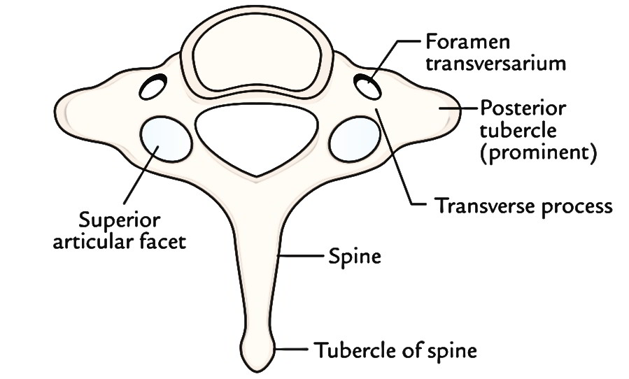
Cervical Vertebrae Drawing - An examination of one of the bones, such as the third cervical vertebra (c3), can be used to show the markings found on the other four. Spinal cord anatomical scheme, vector illustration on white background. Web the atypical vertebrae are cervical level one and two (c1 and c2). 24.1af (a) anatomic drawing of a typical cervical vertebra from a superior projection. Then, begin drawing the vertebra. The c3 vertebra is the first bone of the spinal column to feature the standard vertebral shape, unlike the c1 and c2 vertebrae that. Human spine bones anatomy, vector sketch of skeleton backbone or vertebral column. The c2, the vertebra below it, is also known as the axis. Web blocked or fused vertebrae. Web the vertebrae have laterally directed transverse processes that arise anterior to the facets in the cervical and lumbar spine and posterior to the facets in the thoracic spine.
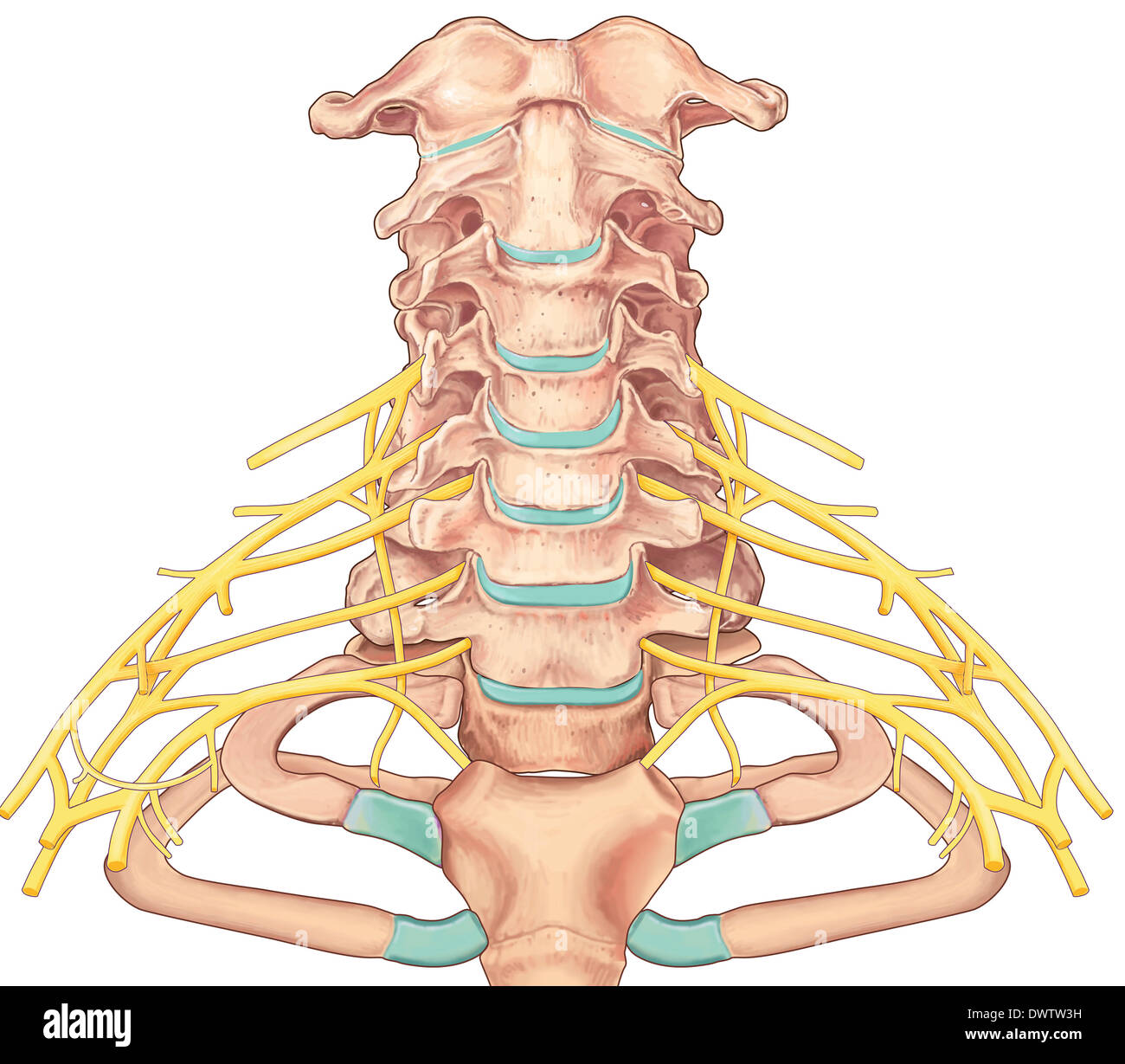
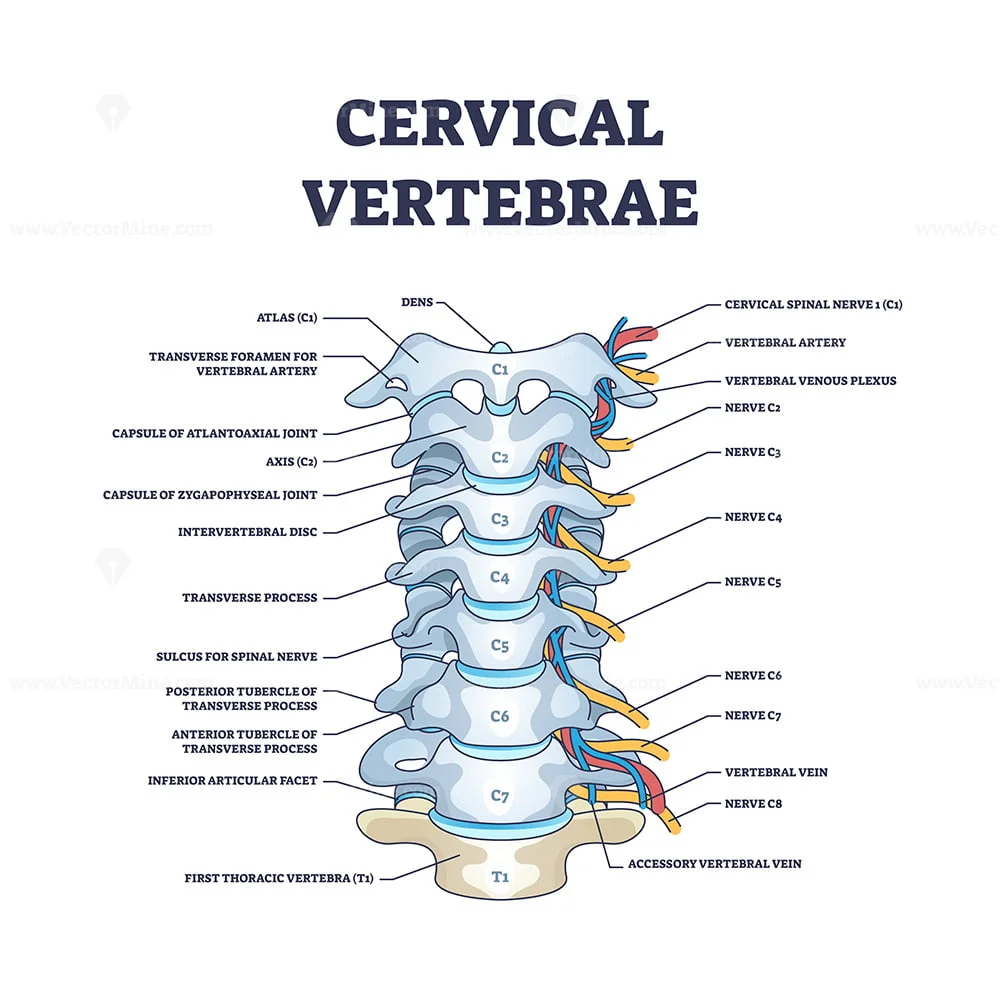
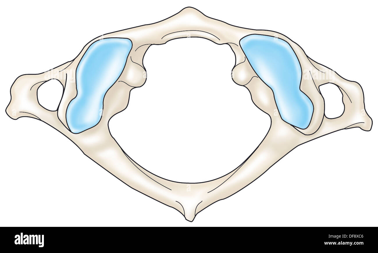
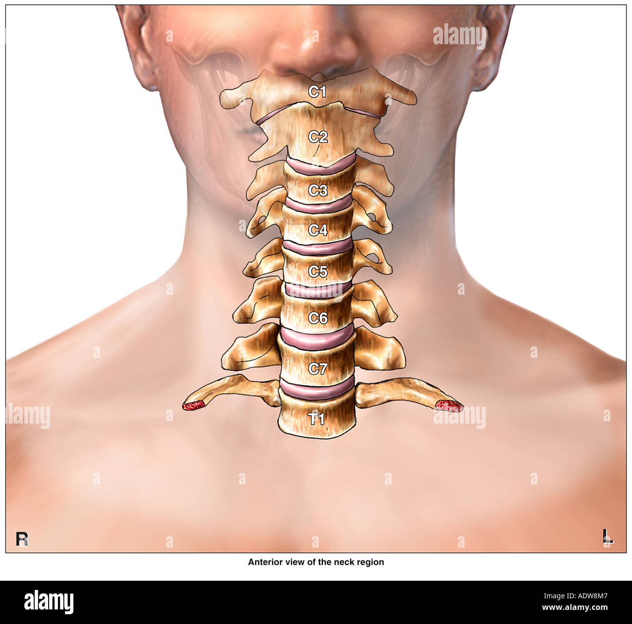
Web the cervical vertebrae are stacked along the length of the neck to form a continuous column between the skull and the chest.mycontentbreak each cervical vertebra is named by its position in order from superior (c1 or first cervical vertebra) to inferior (c7 or seventh cervical vertebra). The first two bones, c1 and c2, are highly specialized, known as the atlas and axis. The third exactly resembles the fourth, and the fifth only differs in a small opening in the lateral arc, indicated in my drawing of the fourth, on the left side. Human spine bones anatomy, vector sketch of skeleton backbone or vertebral column. Cervical vertebrae are found in the neck region, starting from the base of the skull and extending to the thoracic cage of the trunk. Cervical, thoracic and lumbar vertebrae, pelvic curvature and coccyx, rib facet, intervertebral discs and foramen drawing of a cervical spine stock illustrations A cervical spinal cord injury is an injury to your cervical vertebrae. If the patient is supine, this view will allow for all. 24.1af (a) anatomic drawing of a typical cervical vertebra from a superior projection. Use another curved line to complete the outline of the sacrum and the pointed coccyx at its base.
An examination of one of the bones, such as the third cervical vertebra (c3), can be used to show the markings found on the other four. Web the spinal column is divided into three sections, specifically the cervical, thoracic, and lumbar, and it is the uppermost section, the cervical, that makes up the neck. Web all seven cervical vertebrae are numbered. Web the cervical portion of the spine is an important one anatomically and clinically. Most spinal cord injuries are the result of a sudden, traumatic blow to the vertebrae. It is situated inside the vertebral canal of the vertebral column. Use another curved line to complete the outline of the sacrum and the pointed coccyx at its base. It is within this region that the nerves to the arms arise via the brachial plexus, and where the cervical plexus forms providing innervation to the diaphragm among other structures. Spinal cord anatomical scheme, vector illustration on white background. The seventh has no inferior fig.

7th cervical vertebrae coaster medical illustration and Etsy
Each vertebra has a large hole (vertebral foramen) for the spinal. Web the cervical spine performs several crucial roles, including: Protecting the spinal cord.the spinal cord is a bundle of nerves that extends from the brain and runs through the cervical spine and thoracic spine (upper and middle back) prior to ending just before the lumbar spine (lower back). During.
Human Cervical Vertebra Bone ClipArt ETC
A cervical spinal cord injury is an injury to your cervical vertebrae. Web spine bones anatomy, vector sketch of backbone human spine bones anatomy, vector sketch of skeleton backbone or vertebral column. Where are the cervical vertebrae located. This part of the spine is known as the lumbar region. The atlas (c1) consists of two arches (anterior, posterior) and contains.

Cervical vertebrae labeled vector illustration medical diagram Vértebra
Web the cervical vertebrae are stacked along the length of the neck to form a continuous column between the skull and the chest.mycontentbreak each cervical vertebra is named by its position in order from superior (c1 or first cervical vertebra) to inferior (c7 or seventh cervical vertebra). If the patient is supine, this view will allow for all. Use curved.

Cervical vertebra bone graphic hand drawing Vector Image
Web the cervical portion of the spine is an important one anatomically and clinically. The c1, the first vertebra in the column (closest to the skull), is also known as the atlas. The spinal cord is part of the central nervous system (cns). During development, there’s a disproportion between spinal cord growth and vertebral column growth. Cervical one is also.
Cervical vertebra drawing Stock Photo Alamy
An examination of one of the bones, such as the third cervical vertebra (c3), can be used to show the markings found on the other four. Web the spinal column is divided into three sections, specifically the cervical, thoracic, and lumbar, and it is the uppermost section, the cervical, that makes up the neck. The atlas (c1) consists of two.

C II Vertebra Cervicalis Superior view, drawing timelapse. YouTube
Most spinal cord injuries are the result of a sudden, traumatic blow to the vertebrae. Protecting the spinal cord.the spinal cord is a bundle of nerves that extends from the brain and runs through the cervical spine and thoracic spine (upper and middle back) prior to ending just before the lumbar spine (lower back). The c2, the vertebra below it,.
Cervical Vertebrae Earth's Lab
Most spinal cord injuries are the result of a sudden, traumatic blow to the vertebrae. Web the c3 vertebra is a bone of the cervical spine found in the neck around the chin and hyoid bone. The first two bones, c1 and c2, are highly specialized, known as the atlas and axis. Texture the bone with additional curved lines. Accessory.
Cervical vertebrae with bones detailed and labeled structure outline
The c1, the first vertebra in the column (closest to the skull), is also known as the atlas. Texture the bone with additional curved lines. It is situated inside the vertebral canal of the vertebral column. These vertebrae share many anatomical characteristics. It’s third vertebra in the spinal column, inferior to the axis (c2 vertebra) and superior to the c4.
CERVICAL VERTEBRA, DRAWING Stock Photo 61047286 Alamy
Spinal cord anatomical scheme, vector illustration on white background. An examination of one of the bones, such as the third cervical vertebra (c3), can be used to show the markings found on the other four. These vertebrae share many anatomical characteristics. The c2, the vertebra below it, is also known as the axis. It’s third vertebra in the spinal column,.
Anatomy of the Cervical Spine Region showing Neck Vertebrae Stock Photo
The cervical spine also allows passage of important vasculature to reach. The spinal cord is part of the central nervous system (cns). Spinal cord anatomical scheme, vector illustration on white background. Then, begin drawing the vertebra. Use another curved line to complete the outline of the sacrum and the pointed coccyx at its base.
24.1Af (A) Anatomic Drawing Of A Typical Cervical Vertebra From A Superior Projection.
Hand drawn vector spine isolated on white. Where are the cervical vertebrae located. Use another curved line to complete the outline of the sacrum and the pointed coccyx at its base. The c3 vertebra is the first bone of the spinal column to feature the standard vertebral shape, unlike the c1 and c2 vertebrae that.
The C1, Or First Cervical Vertebra, Is Commonly Called The Atlas Due To Its Unique Position In The Spine.
Web the cervical spine performs several crucial roles, including: An examination of one of the bones, such as the third cervical vertebra (c3), can be used to show the markings found on the other four. Most spinal cord injuries are the result of a sudden, traumatic blow to the vertebrae. If the patient is supine, this view will allow for all.
Texture The Bone With Additional Curved Lines.
The seventh has no inferior fig. Web all seven cervical vertebrae are numbered. Spinal cord anatomical scheme, vector illustration on white background. Web #biology #typicalcervicalvertebra #diagramofcervicalvertebra #cervicalvetebra #class11 #hscbiology #maharashtrastateboard2021 #goaboard2021 #biology2021 #bio.
(B) Anatomic Drawing Of A Typical Cervical Vertebra From A Lateral.
The third exactly resembles the fourth, and the fifth only differs in a small opening in the lateral arc, indicated in my drawing of the fourth, on the left side. Web the atypical vertebrae are cervical level one and two (c1 and c2). Then, begin drawing the vertebra. In greek mythology, atlas was the titan who held the earth on his shoulders, just like the atlas holds the skull on top of the neck.mycontentbreak the atlas is located at the top of the neck, just inferior to the condyles of the.