Cilia Drawing
Cilia Drawing - 73k views 6 years ago cells. This is the well labelled diagram. Web biology (kimball) unit 3: Web choose from drawing of cilia stock illustrations from istock. If only one, or a few, they are flagella. Web 📝 find notes here: Web here, we provide an introductory overview of our current understanding of the structure and function of the cilium, with a focus on the signaling pathways that are coordinated by primary cilia to ensure proper organ generation and maintenance. These whiplike appendages extend from the surface of many types of eukaryotic cells. Cilia can be divided into two types: Whereas primary cilia have relatively little additional structure,.
When present, the cell has just one flagellum or a few flagella. So friends if you have problem in any other diagram so tell me in comment box. This video is regarding how to draw section of cilia or flagella. This is the well labelled diagram. Web here, we provide an introductory overview of our current understanding of the structure and function of the cilium, with a focus on the signaling pathways that are coordinated by primary cilia to ensure proper organ generation and maintenance. They are also involved in mechanoreception. Web choose from drawing of a cilia stock illustrations from istock. These appendages have been studied in different groups of animals for their functions of both movement and sensation as they can detect changes in the environmental composition and ph. Cilia, primary cilium, ciliopathies, signaling pathways, syndromic diseases. Long black lashes vector illustration.
Web 2.6k views 1 year ago science diagrams | explained and labelled science diagrams. Web choose from drawing of cilia stock illustrations from istock. Web choose from cilia drawing stock illustrations from istock. Cilia, primary cilium, ciliopathies, signaling pathways, syndromic diseases. Cilia play a major role in locomotion. The cellular basis of life. This video is regarding how to draw section of cilia or flagella. Web in these 3d images of epithelial cells in a mouse’s trachea, scientist and johns hopkins university president’s frontier award winner andrew holland and ph.d. They are primitive in nature and could be single or many. When present, the cell has just one flagellum or a few flagella.
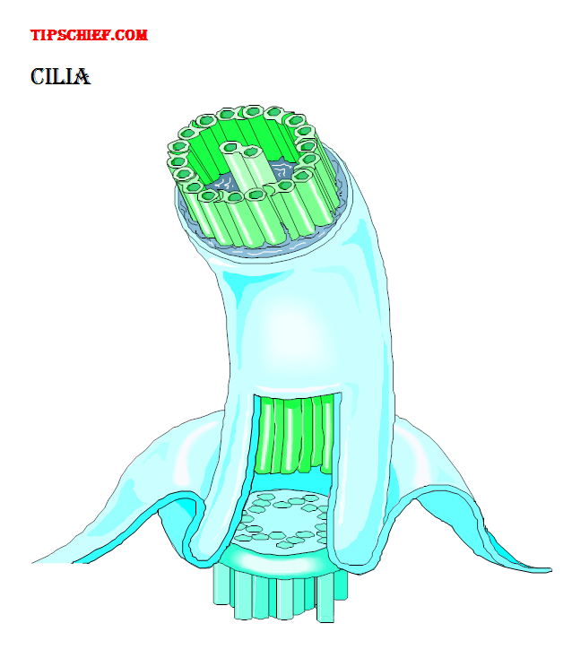
diagram of cilia of biology images
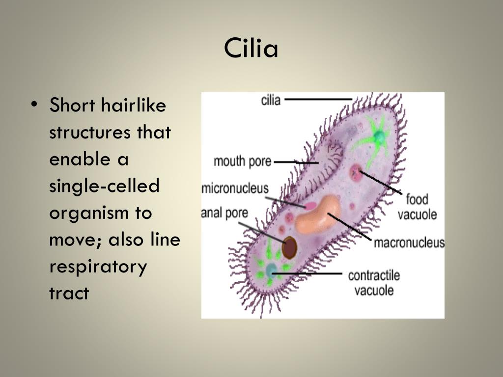
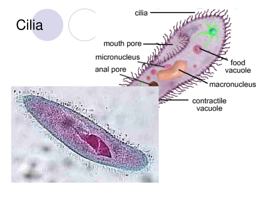
They are primarily responsible for locomotion, either of the cell itself or of fluids on the cell surface. They are also involved in mechanoreception. If only one, or a few, they are flagella. Web in these 3d images of epithelial cells in a mouse’s trachea, scientist and johns hopkins university president’s frontier award winner andrew holland and ph.d. Candidate gina.
Cilia structure Science, Biology ShowMe
How to draw a diagram of structure of cilia or flagella in exam is the topic. Candidate gina lomastro demonstrate how cells build cilia. They are primarily responsible for locomotion, either of the cell itself or of fluids on the cell surface. Web schematics of cilia/flagella (green) in the ciliated protozoa tetrahymena (a), the alga chlamydomonas (b), the caenorhabditis elegans.

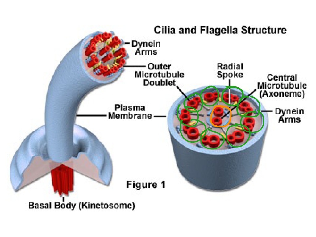
Cilia structure. Left the ciliary axoneme is composed by nine
Web schematics of cilia/flagella (green) in the ciliated protozoa tetrahymena (a), the alga chlamydomonas (b), the caenorhabditis elegans sensory neuron adl (c), a mammalian rod photoreceptor (d), a mammalian hypothalamic neuron (e), and a mammalian airway epithelial cell (f). So friends if you have problem in any other diagram so tell me in comment box. Drawings are not to scale..

Illustration of human cilia Stock Image F023/5120 Science Photo
They are primitive in nature and could be single or many. If there are many of them, they are called cilia. The cellular basis of life. These whiplike appendages extend from the surface of many types of eukaryotic cells. Web see how coral organisms sweep water into turbulent patterns with their cilia, drawing in nutrients and expelling wastes.
Animal Cell Cilia
Web choose from cilia drawing stock illustrations from istock. If there are many of them, they are called cilia. These whiplike appendages extend from the surface of many types of eukaryotic cells. 73k views 6 years ago cells. Web 📝 find notes here:

How to Draw Section of cilia or flagella Structure of Cilia or
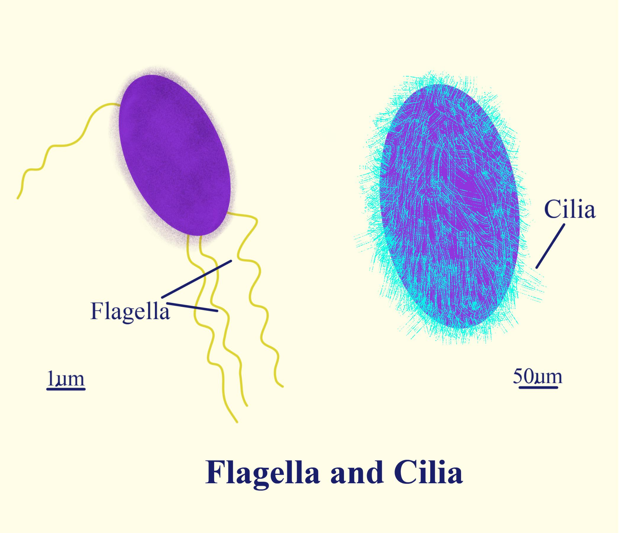
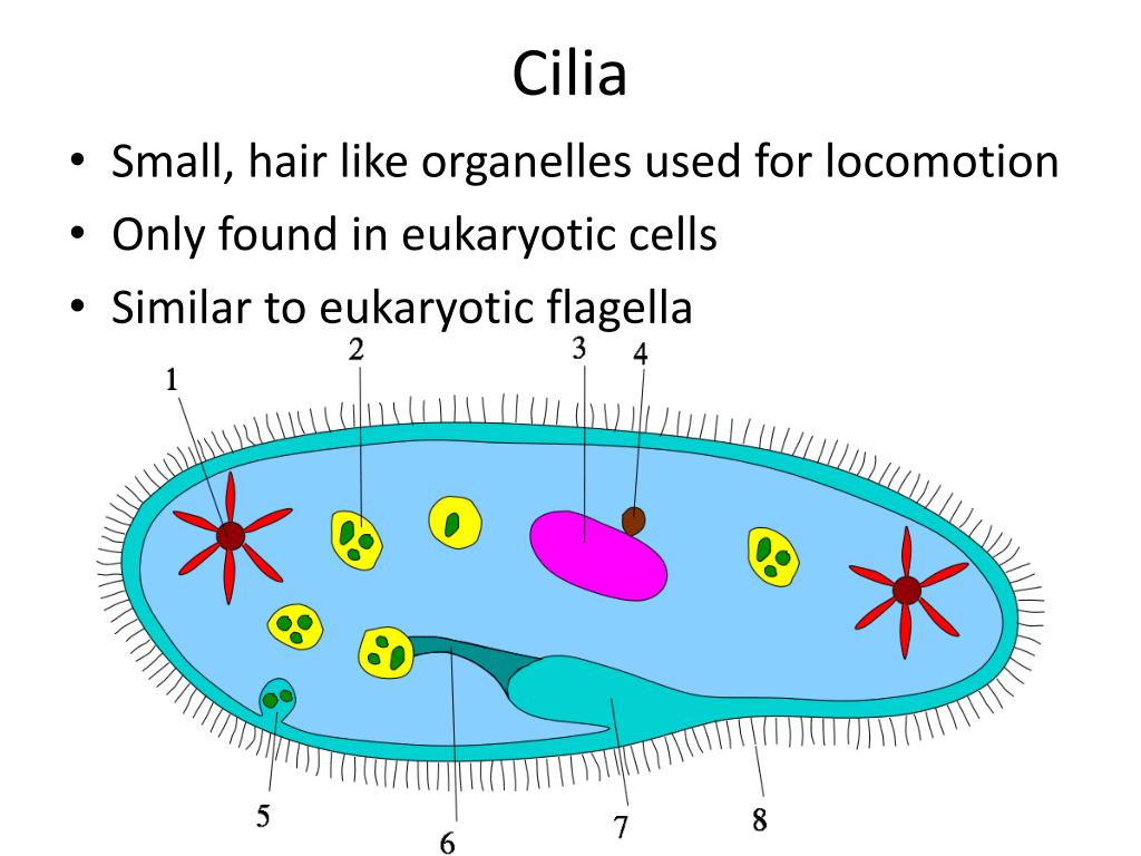
Web diagram of ciliary structure. If there are many of them, they are called cilia. Web compare and contrast cilia and flagella; This is the well labelled diagram. Web see how coral organisms sweep water into turbulent patterns with their cilia, drawing in nutrients and expelling wastes.

Structure of the primary cilium. The core structure of primary cilia is
When present, the cell has just one flagellum or a few flagella. Cilia drawing pictures, images and stock photos. Web diagram of ciliary structure. This video is regarding how to draw section of cilia or flagella. Candidate gina lomastro demonstrate how cells build cilia.
What Is Cilia In Biology? Body Only Now
Web see how coral organisms sweep water into turbulent patterns with their cilia, drawing in nutrients and expelling wastes. Candidate gina lomastro demonstrate how cells build cilia. Web it is very easy to draw. If only one, or a few, they are flagella. Web 2.6k views 1 year ago science diagrams | explained and labelled science diagrams.
Animal Cell Cilia
Whereas primary cilia have relatively little additional structure,. When present, the cell has just one flagellum or a few flagella. Drawings are not to scale. Prokaryotes sometimes have flagella, but they are structurally very different from eukaryotic flagella. These whiplike appendages extend from the surface of many types of eukaryotic cells.
PPT Cilia and Flagella in Cell Structure PowerPoint Presentation
They are also involved in mechanoreception. Web compare and contrast cilia and flagella; Web diagram of ciliary structure. Summarize the differences among the components of prokaryotic cells, animal cells, and plant cells This video is regarding how to draw section of cilia or flagella.
The Cellular Basis Of Life.
The image shows red, elongated structures, which are cilia. Web choose from cilia drawing stock illustrations from istock. Drawings are not to scale. So friends if you have problem in any other diagram so tell me in comment box.
Web In These 3D Images Of Epithelial Cells In A Mouse’s Trachea, Scientist And Johns Hopkins University President’s Frontier Award Winner Andrew Holland And Ph.d.
Prokaryotes sometimes have flagella, but they are structurally very different from eukaryotic flagella. Web the cilium (plural: Whereas primary cilia have relatively little additional structure,. This video is regarding how to draw section of cilia or flagella.
Web Here, We Provide An Introductory Overview Of Our Current Understanding Of The Structure And Function Of The Cilium, With A Focus On The Signaling Pathways That Are Coordinated By Primary Cilia To Ensure Proper Organ Generation And Maintenance.
If only one, or a few, they are flagella. Web biology (kimball) unit 3: If there are many of them, they are called cilia. Web diagram of ciliary structure.
When Present, The Cell Has Just One Flagellum Or A Few Flagella.
They are primitive in nature and could be single or many. Cilia play a major role in locomotion. Web it is very easy to draw. This is the well labelled diagram.