Draw And Label A Sarcomere
Draw And Label A Sarcomere - Include the myofilaments, but do not include the regulatory proteins that turn on and off muscle contraction, or the names of the light and. Mainly of actin and myosin proteins. This is an online quiz called sarcomere labeling. The sarcomere is the functional (contractile) unit of skeletal muscle. 1.4k views 2 years ago science diagrams | explained and labelled science diagrams. So, there is a need arise for a unit which can repay for the. How to draw a diagram of diagram of sarcomere/showing i band, a band, h zone and. The actin filaments radiate out from the z discs and help to. Actin and the z discs are shown in red. Web the sarcomere structure is so crucial in this theory because a muscle needed to physically shorten.
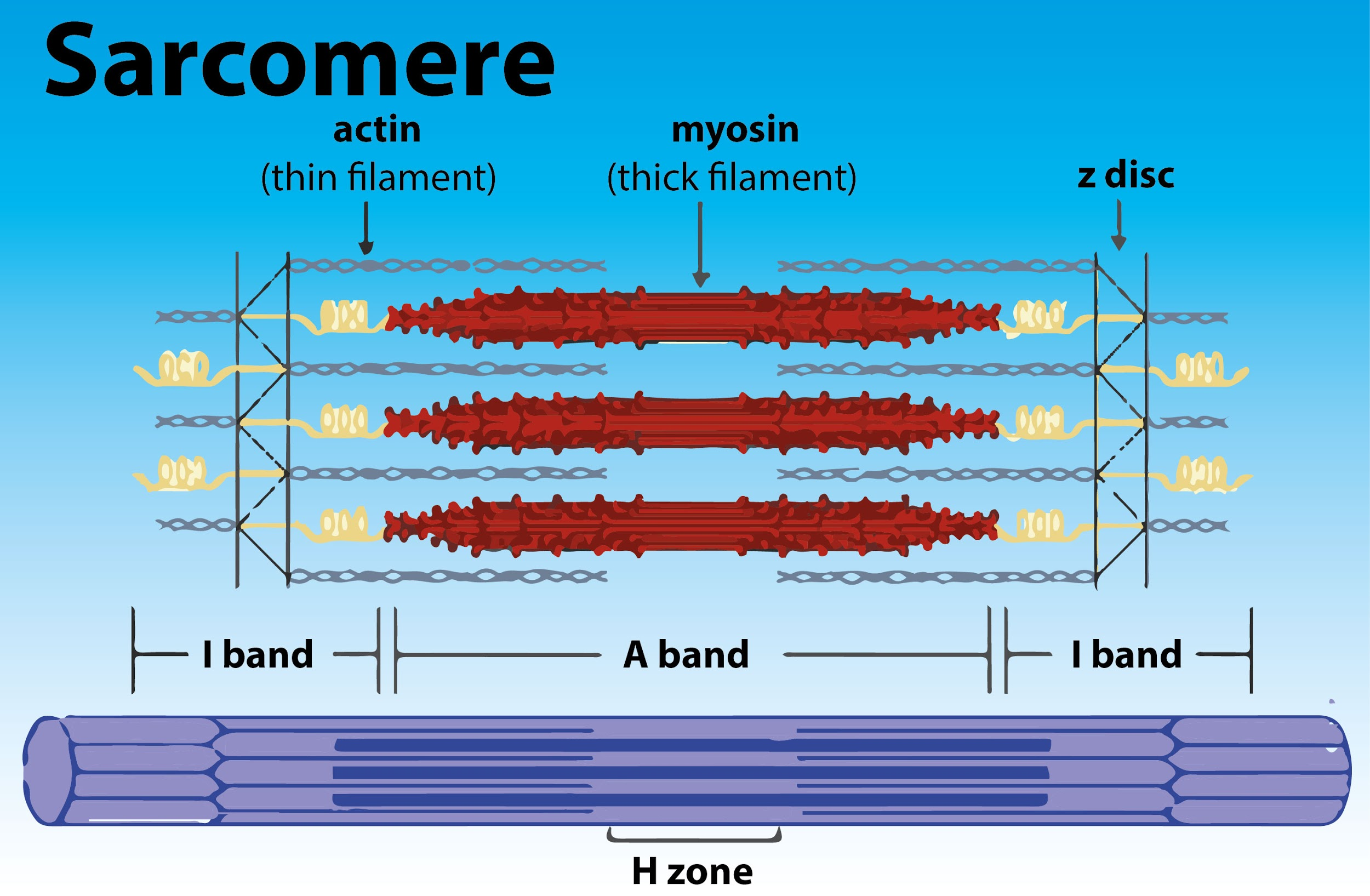
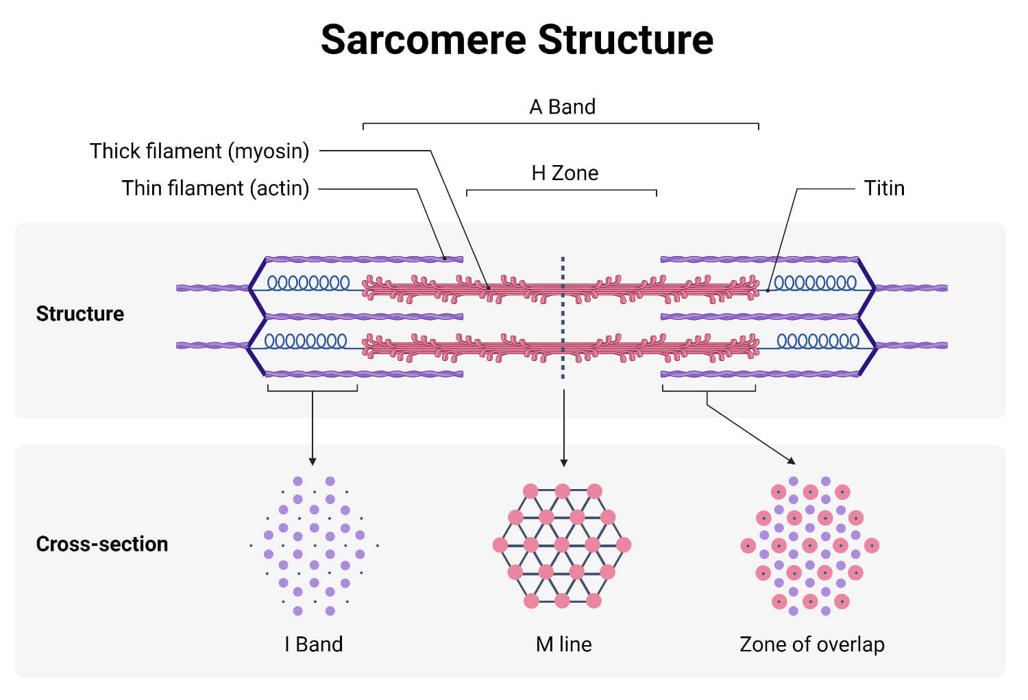
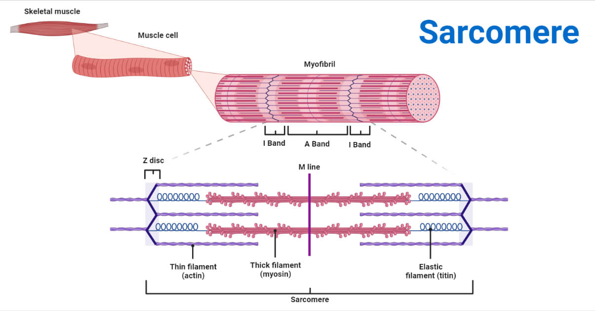
You'll get a detailed solution from a subject. Web (a) the basic organization of a sarcomere subregion, showing the centralized location of myosin (a band). Web each individual sarcomere is flanked by dense protein discs called z lines, which hold the myofilaments in place. The sarcomere fundamentally consists of two main myofilaments: Web sarcomere labeling — quiz information. Draw and label a picture of a sarcomere. Thick filaments called myosin and thin. This is an online quiz called sarcomere labeling. The actin filaments radiate out from the z discs and help to. You can use it as sarcomere labeling practice, completely free to.
The thick filament is composed of the. The diagrammatic representation of a sarcomere is as follows: Web each individual sarcomere is flanked by dense protein discs called z lines, which hold the myofilaments in place. Web (a) the basic organization of a sarcomere subregion, showing the centralized location of myosin (a band). This problem has been solved! Include actin, myosin, the z lines, the h zone, and the i band. You can use it as sarcomere labeling practice, completely free to. It is the region of a myofibril between two z discs. Learn vocabulary, terms, and more with flashcards, games, and other study tools. Web a sarcomere is a microscopic segment repeating in a myofibril.
Draw the diagram of a of skeletal muscle showing different
The actin filaments radiate out from the z discs and help to. This is an online quiz called sarcomere labeling. This problem has been solved! Learn everything about its anatomy and structure on kenhub! Web sarcomere labeling — quiz information.
[Solved] 12. Draw and label the parts of a Course Hero
The diagrammatic representation of a sarcomere is as follows: Web (a) the basic organization of a sarcomere subregion, showing the centralized location of myosin (a band). The sarcomere fundamentally consists of two main myofilaments: Web start studying label the sarcomere structure. This is an online quiz called sarcomere labeling.

Schematic of structure. are the functional units
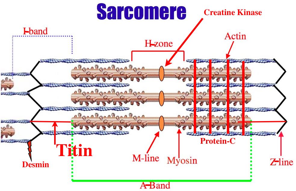
Sarcomeres are contractile units of skeletal muscle that divide into “i” and “a” bands, “m” and “z” lines, and the “h” zone. Draw and label a relaxed sarcomere. Learn vocabulary, terms, and more with flashcards, games, and other study tools. Mainly of actin and myosin proteins. This problem has been solved!.
Definition, Structure, Diagram, and Functions
This problem has been solved!. Include actin, myosin, the z lines, the h zone, and the i band. Web the sarcomere is a main contractile unit of muscle fiber in the skeletal muscle. The sarcomere is the functional (contractile) unit of skeletal muscle. Label the muscles of the face.
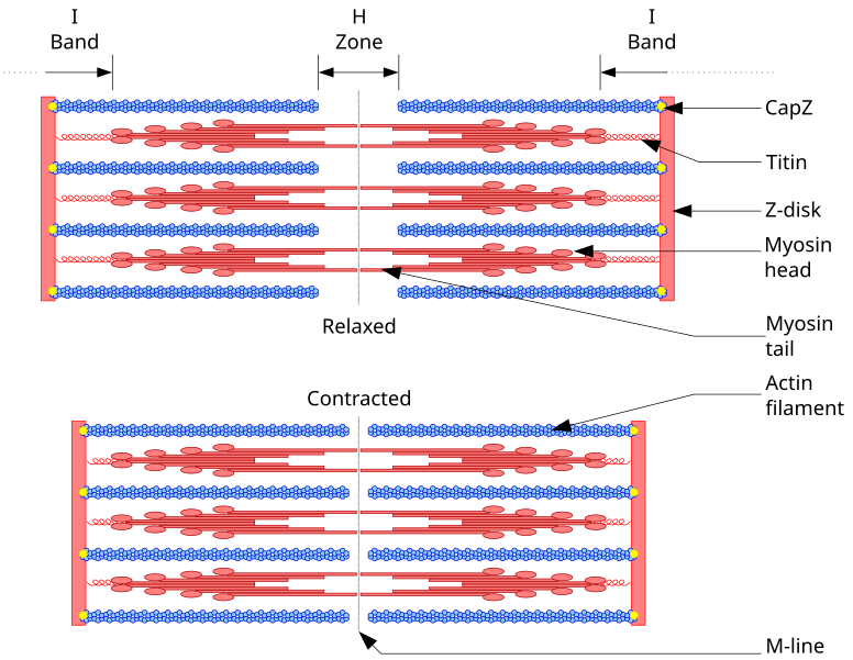
Diagram Of A
Learn vocabulary, terms, and more with flashcards, games, and other study tools. You can use it as sarcomere labeling practice, completely free to. Label the muscles of the face. Web each individual sarcomere is flanked by dense protein discs called z lines, which hold the myofilaments in place. Web (a) the basic organization of a sarcomere subregion, showing the centralized.

10.2 Skeletal Muscle Anatomy and Physiology
This is an online quiz called sarcomere labeling. The actin filaments radiate out from the z discs and help to. 1.4k views 2 years ago science diagrams | explained and labelled science diagrams. Draw and label a picture of a sarcomere. Actin and the z discs are shown in red.
anatomy of a
Anatomy and physiology questions and answers. The sarcomere fundamentally consists of two main myofilaments: How to draw a diagram of diagram of sarcomere/showing i band, a band, h zone and. The sarcomere is the functional (contractile) unit of skeletal muscle. The actin filaments radiate out from the z discs and help to.

Diagram Diagram Quizlet
1.4k views 2 years ago science diagrams | explained and labelled science diagrams. Web the sarcomere is a main contractile unit of muscle fiber in the skeletal muscle. Label the muscles of the face. Web the sarcomere structure is so crucial in this theory because a muscle needed to physically shorten. The actin filaments radiate out from the z discs.
Definition, Structure, Diagram, and Functions
Actin and the z discs are shown in red. Learn everything about its anatomy and structure on kenhub! The thick filament is composed of the. How to draw a diagram of diagram of sarcomere/showing i band, a band, h zone and. The sarcomere is the functional (contractile) unit of skeletal muscle.

muscular biology scheme vector illustration VectorMine
Learn vocabulary, terms, and more with flashcards, games, and other study tools. Learn everything about its anatomy and structure on kenhub! Draw and label a picture of a sarcomere. Sarcomeres are contractile units of skeletal muscle that divide into “i” and “a” bands, “m” and “z” lines, and the “h” zone. The thick filament is composed of the.
Web Start Studying Label The Sarcomere Structure.
You can use it as sarcomere labeling practice, completely free to. Mainly of actin and myosin proteins. Thick filaments called myosin and thin. This problem has been solved!
Draw And Label A Picture Of A Sarcomere.
Web the sarcomere is a main contractile unit of muscle fiber in the skeletal muscle. Label the muscles of the face. The thick filament is composed of the. The diagrammatic representation of a sarcomere is as follows:
Learn Everything About Its Anatomy And Structure On Kenhub!
Web (a) the basic organization of a sarcomere subregion, showing the centralized location of myosin (a band). 1.4k views 2 years ago science diagrams | explained and labelled science diagrams. This problem has been solved!. Draw and label a relaxed sarcomere.
The Sarcomere Is The Functional (Contractile) Unit Of Skeletal Muscle.
Web sarcomere labeling — quiz information. The actin filaments radiate out from the z discs and help to. Web a sarcomere is a highly organized structure made up of thick and thin protein filaments; Anatomy and physiology questions and answers.