Draw And Label Microscope
Draw And Label Microscope - In this tutorial, writing master shows you how to. Web labeled parts of a microscope. In this interactive, you can label the different parts of a microscope. The eyepiece usually contains a 10x or 15x power lens. December 14, 2022 by ramzan asghar. Ready to take your drawing skills to the next level? This activity has been designed for use in homes and schools. All microscopes share features in common. Eyepieces typically have a magnification between 5x & 30x. There are six printables available.
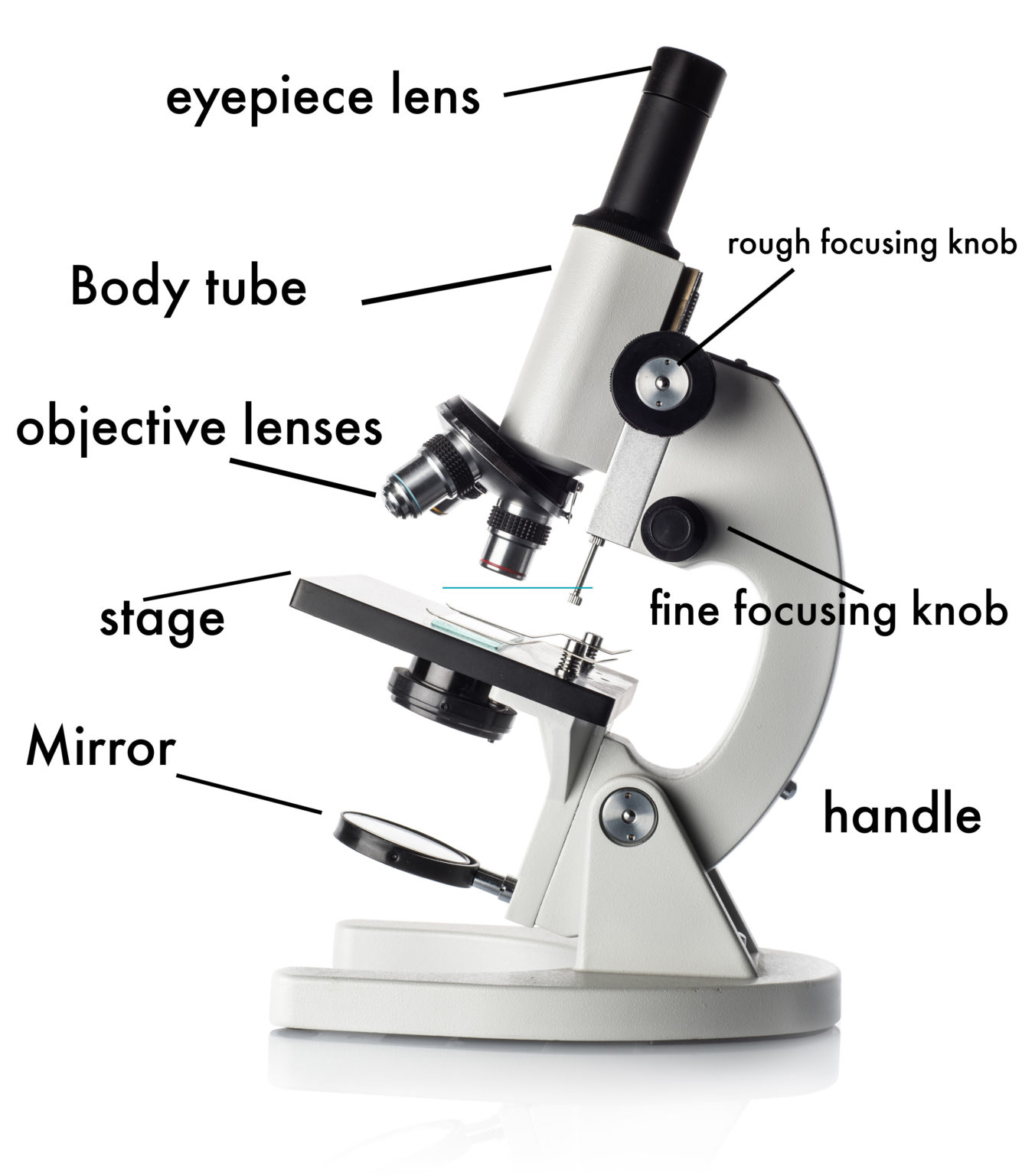
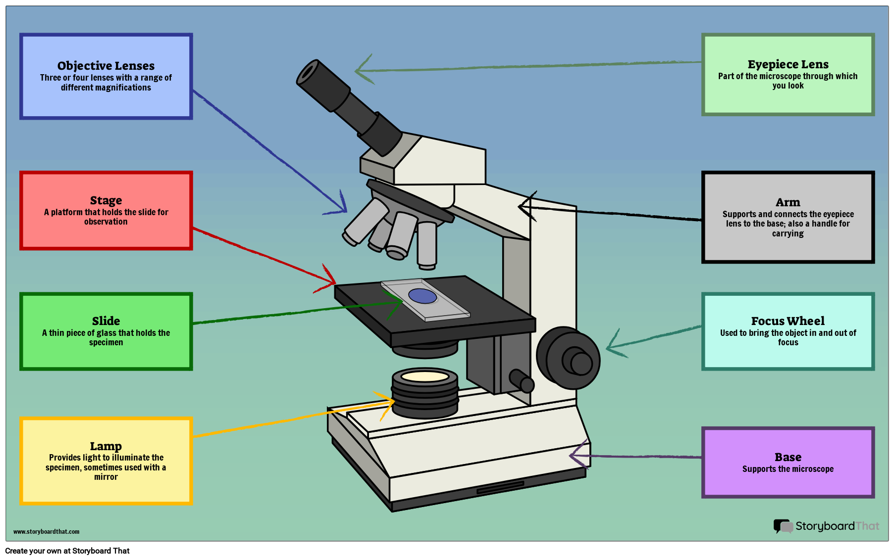
Web parts of a microscope with labeled diagram and functions. In this tutorial, writing master shows you how to. All microscopes share features in common. Optical components of a compound microscope. Web the secret to drawing an excellent microscope picture is attention to detail and familiarity with the structure and components. The most important feature of a microscope is that it magnifies the image of the specimen and resolves it better to help the observers see the specimen clearly to carry out their experiments. First and foremost, we have a labeled microscope diagram, available in both black and white and color. Never turn the nose piece by the objective lens. Label the cell wall, cell membrane, cytoplasm, and chloroplasts in your lab manual. Structural parts of a microscope and their functions.
The lens the viewer looks through to see the specimen. Use only lens paper to clean microscope lenses. The body tube connects the eyepiece to the objective lenses. Web use this interactive to identify and label the main parts of a microscope. #microscope #howtodraw #adimushow this is an easy and simple drawing of microscope. 6.4k views 1 year ago #microscope #howtodraw #adimushow. Label the parts of the microscope with answers (a4) pdf print version. Explain or describe the parts of a light compound microscope. The labeling worksheet could be used as a quiz or as part of direct instruction. Useful as a study guide for learning the anatomy of a microscope.

Simple Microscope Definition, Principle, Magnification, Parts
Useful as a study guide for learning the anatomy of a microscope. Before starting, make sure you have all the necessary materials handy. Also indicate the estimated cell size in micrometers under your drawing. 6.4k views 1 year ago #microscope #howtodraw #adimushow. The eyepiece usually contains a 10x or 15x power lens.

Parts of a microscope with functions and labeled diagram
Structural parts of a microscope and their functions. Major structural parts of a compound microscope. The most important feature of a microscope is that it magnifies the image of the specimen and resolves it better to help the observers see the specimen clearly to carry out their experiments. Before starting, make sure you have all the necessary materials handy. All.
How to Use a Microscope
This simple worksheet pairs with a lesson on the light microscope, where beginning biology students learn the parts of the light microscope and the steps needed to focus a slide under high power. Eyepieces typically have a magnification between 5x & 30x. This activity has been designed for use in homes and schools. Web labeling the parts of the microscope.
36+ Label Each Part Of A Microscope Gif Diagram Printabel
Explain or describe the parts of a light compound microscope. A microscope is an essential tool for scientists, researchers, and medical professionals. The part that is looked through at the top of the compound microscope. The eyepiece usually contains a 10x or 15x power lens. Web labeled diagram of a compound microscope.

Labeled Microscope Diagram Tim's Printables
Structural support that holds & connects the eyepieces to the objective lenses. In this tutorial, writing master shows you how to. To use a light microscope to observe, draw and label a selection of plant and animal cells, including a magnification scale. The most important feature of a microscope is that it magnifies the image of the specimen and resolves.

How to Draw a Microscope and Label Nesecale Thiptin
Optical components of a compound microscope. Also indicate the estimated cell size in micrometers under your drawing. First and foremost, we have a labeled microscope diagram, available in both black and white and color. In other words, it enlarges images of small objects. Web learn about the different parts of the microscope, including the simple microscope and the compound microscope,.

Simple Microscope Drawing at GetDrawings Free download
The part that is looked through at the top of the compound microscope. In other words, it enlarges images of small objects. In this interactive, you can label the different parts of a microscope. Web compound microscope definitions for labels. Before starting, make sure you have all the necessary materials handy.

Microscope diagram Tom Butler Technical Drawing and Illustration
To use a light microscope to observe, draw and label a selection of plant and animal cells, including a magnification scale. #microscope #howtodraw #adimushow this is an easy and simple drawing of microscope. Never turn the nose piece by the objective lens. Major structural parts of a compound microscope. Invented by a dutch spectacle maker in the late 16th century,.

How To Draw A Microscope 🔬 YouTube
A microscope has two sets of lenses. The microscope layout, including the blank and answered versions are available as pdf downloads. Web the secret to drawing an excellent microscope picture is attention to detail and familiarity with the structure and components. The most important feature of a microscope is that it magnifies the image of the specimen and resolves it.

Microscope Diagram Labeled, Unlabeled and Blank Parts of a Microscope
453 views 3 years ago #labels #chatgpt #drawing. Coarse and fine focus knobs. This activity has been designed for use in homes and schools. Web labeling the parts of the microscope | microscope world resources. Web compound microscope definitions for labels.
A Microscope Is An Essential Tool For Scientists, Researchers, And Medical Professionals.
After completing the lab, the student will be able to: Optical components of a compound microscope. Drag and drop the text labels onto the microscope diagram. Web overview of microscope and diagram.
What Do You Think Is The Purpose Of Each Lens?
The body tube connects the eyepiece to the objective lenses. Use this with the microscope parts activity to help students identify and label the main parts of. Structural support that holds & connects the eyepieces to the objective lenses. Label the parts of the microscope (a4) pdf print version.
Major Structural Parts Of A Compound Microscope.
Web labeled parts of a microscope. The most important feature of a microscope is that it magnifies the image of the specimen and resolves it better to help the observers see the specimen clearly to carry out their experiments. Explain or describe the parts of a light compound microscope. Web use this interactive to identify and label the main parts of a microscope.
Structural Parts Of A Microscope And Their Functions.
Never turn the nose piece by the objective lens. Eyepieces typically have a magnification between 5x & 30x. In this tutorial, writing master shows you how to. Eyepiece (ocular lens) with or without pointer: