Draw Diagram Of Heart
Draw Diagram Of Heart - Your ribcage protects your heart, everyone’s heart is a slightly. Electrical impulses make your heart beat, moving blood through these chambers. Web practise labelling the human heart diagram. At the heart of it all: In coordination with valves, the chambers work to keep blood flowing in the proper sequence. The heart wall is made up of three layers: Anatomical illustrations and structures, 3d model and photographs of dissection. Then, fill in the base of the heart with the right and left ventricles and the right and left atriums. [1] the main shape will be the basis for the left and right ventricles. How to draw human heart step by step!
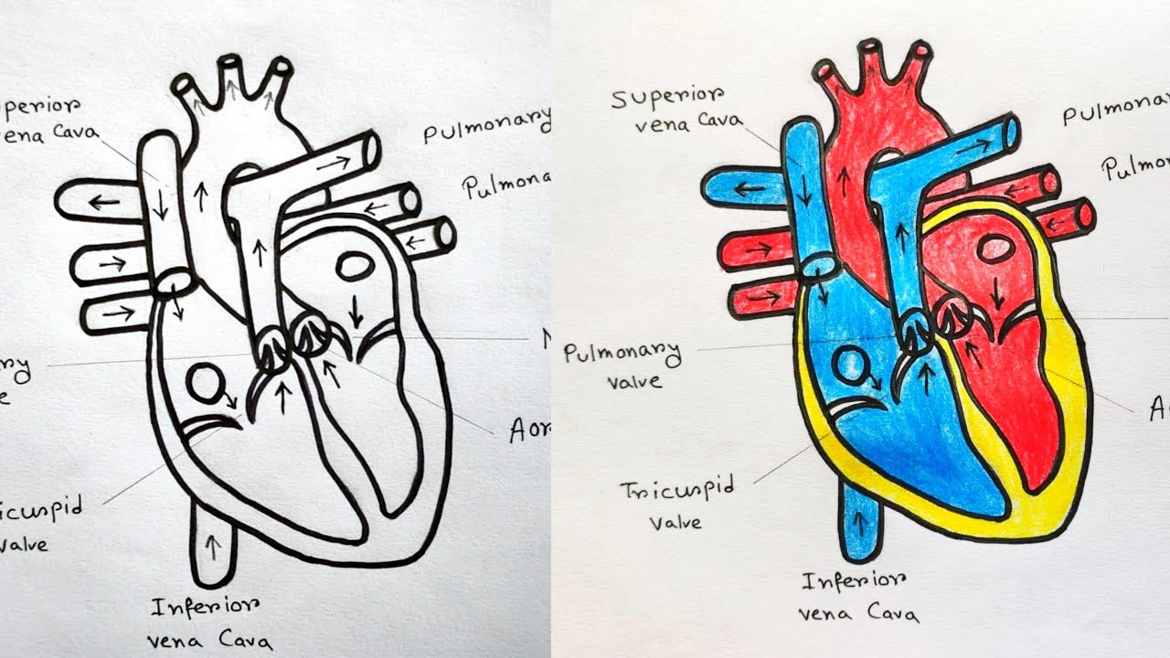
The lower two chambers of the heart are called ventricles. July 25, 2023 fact checked. It also has several margins: The heart has five surfaces: Anatomy and function of the heart. 1.1m views 3 years ago drawing tutorials. This outlines the lower chamber of the heart, which includes both the left and right ventricles. In coordination with valves, the chambers work to keep blood flowing in the proper sequence. Web your heart is located in the front of your chest. Web to draw the internal structure of the heart, start by sketching the 2 pulmonary veins to the lower left of the aorta and the bottom of the inferior vena cava slightly to the right of that.
Includes an exercise, review worksheet, quiz, and model drawing of an anterior vi The heart is a muscular organ that pumps blood through the blood vessels of the circulatory system. Electrical impulses make your heart beat, moving blood through these chambers. Your heart contains four muscular sections ( chambers) that briefly hold blood before moving it. Your brain and nervous system direct your heart’s function. The heart is made up of four chambers: Introduction to the human heart. The outer layer of the heart wall is called epicardium. A thin layer of tissue, the pericardium, covers the outside, and another layer, the endocardium, lines the inside. Anatomy and function of the heart.

How to Draw the Internal Structure of the Heart 13 Steps
Web medically reviewed by the healthline medical network — by the healthline editorial team — updated on january 20, 2018. Blood transports oxygen and nutrients to the body. 1.1m views 3 years ago drawing tutorials. The upper two chambers of the heart are called auricles. Web function and anatomy of the heart made easy using labeled diagrams of cardiac structures.

human heart drawing labeled
The heart is a mostly hollow, muscular organ composed of cardiac muscles. 324k views 1 year ago human body parts, skeleton& organs. The heart is a muscular organ that pumps blood through the blood vessels of the circulatory system. Within the triangle, draw a horizontal and vertical centerline to split the triangle into four pieces. Web create a curved shape.
Heart Diagram Clipart at GetDrawings Free download
Your ribcage protects your heart, everyone’s heart is a slightly. The middle layer of the heart wall is called myocardium. It should look a bit like the shape of africa. Web to draw the internal structure of the heart, start by sketching the 2 pulmonary veins to the lower left of the aorta and the bottom of the inferior vena.
When one teaches, two learn. The heart and the circulatory system
Written by kelly medford | edited by carmine shannon. It should look a bit like the shape of africa. It is also involved in the removal of. Web in this lecture, dr mike shows the two best ways to draw and label the heart! The lower two chambers of the heart are called ventricles.
Anatomical Drawing Heart at GetDrawings Free download
The user can show or hide the anatomical labels which provide a useful tool to create illustrations perfectly adapted for teaching. Anatomical illustrations and structures, 3d model and photographs of dissection. The size of the heart is the size of about a clenched fist. Web your heart is located in the front of your chest. July 25, 2023 fact checked.
How to draw Human Heart with colour Human Heart labelled diagram
How to draw human heart diagram easily/ human heart diagram drawing in this video i used artline shading pencil and. 1.1m views 3 years ago drawing tutorials. Web practise labelling the human heart diagram. It is a muscular organ with four chambers. Draw the main shape of your human heart drawing.
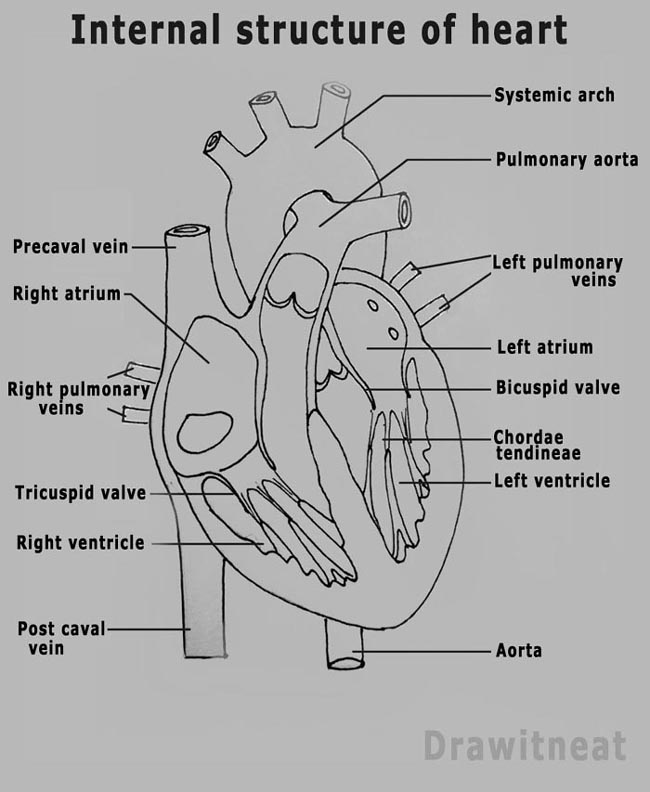
DRAW IT NEAT How to draw human heart labeled
The middle layer of the heart wall is called myocardium. Then, fill in the base of the heart with the right and left ventricles and the right and left atriums. Introduction to the human heart. This outlines the lower chamber of the heart, which includes both the left and right ventricles. Web your heart is located in the front of.
Human Heart Drawing Simple at Explore collection
The human heart is one of the most important organs responsible for sustaining life. Base (posterior), diaphragmatic (inferior), sternocostal (anterior), and left and right pulmonary surfaces. The heart has five surfaces: Written by kelly medford | edited by carmine shannon. Draw the main shape of your human heart drawing.
Heart And Labels Drawing at GetDrawings Free download
It is a muscular organ with four chambers. At the heart of it all: This outlines the lower chamber of the heart, which includes both the left and right ventricles. 406k views 1 year ago. [1] the main shape will be the basis for the left and right ventricles.

How to Draw the Internal Structure of the Heart 14 Steps
The human heart is one of the most important organs responsible for sustaining life. Then, fill in the base of the heart with the right and left ventricles and the right and left atriums. The inferior tip of the heart, known as the apex, rests just superior to the diaphragm. Rotate the 3d model to see how the heart's valves.
July 25, 2023 Fact Checked.
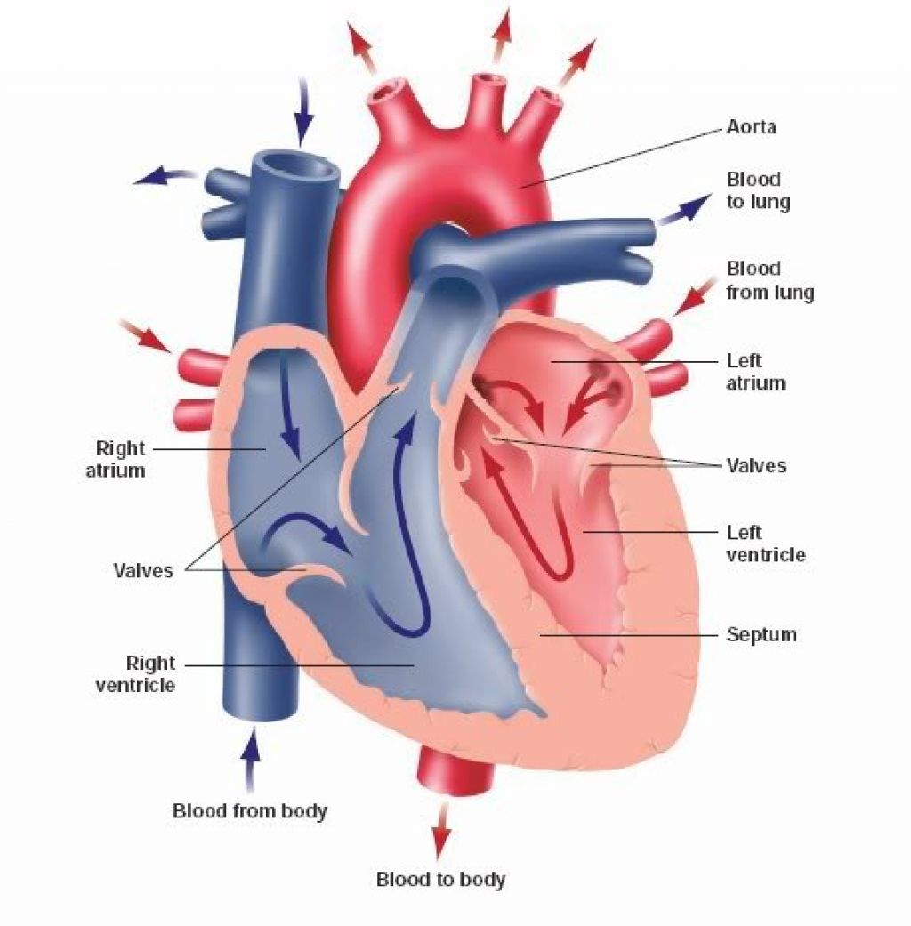
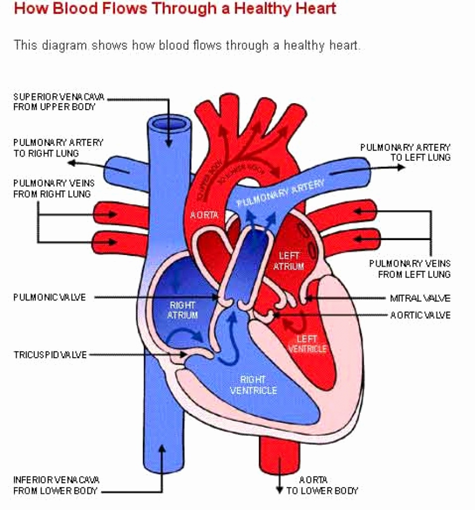
324k views 1 year ago human body parts, skeleton& organs. Web using a simple diagram to show the order in which blood flows through the heart, we will walk through the cardiac circulation pathway in 12 simple steps. It also has several margins: A thin layer of tissue, the pericardium, covers the outside, and another layer, the endocardium, lines the inside.
The Heart Is A Mostly Hollow, Muscular Organ Composed Of Cardiac Muscles.
The middle layer of the heart wall is called myocardium. Blood transports oxygen and nutrients to the body. As with every ezmed post, we have some simple tricks and charts that will help you remember the anatomy, physiology, and function of the right and left side of the heart. Within the triangle, draw a horizontal and vertical centerline to split the triangle into four pieces.
Draw The First Construction Lines.
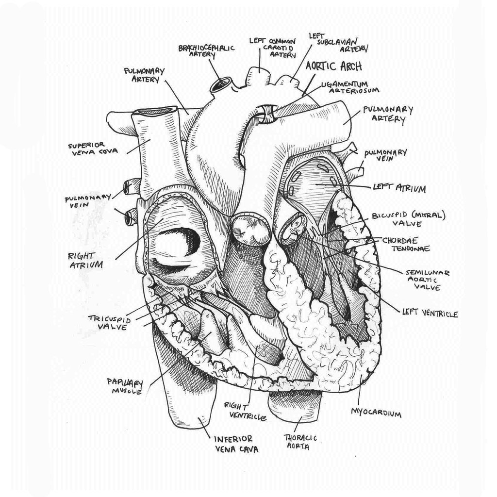
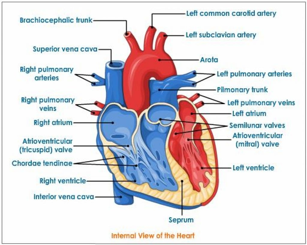
On its superior end, the base of the heart is attached to the aorta,mycontentbreak pulmonary arteries and veins, and the vena cava. The human heart is one of the most important organs responsible for sustaining life. Web this interactive atlas of human heart anatomy is based on medical illustrations and cadaver photography. The size of the heart is the size of about a clenched fist.
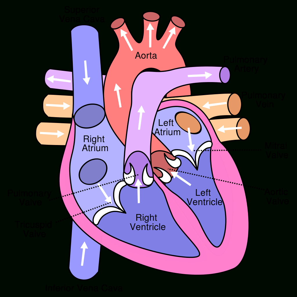
The Heart Cavity Is Divided Down The Middle Into A Right And A Left Heart, Which In Turn Are Subdivided Into Two Chambers.
Right, left, superior, and inferior: Anatomy and function of the heart. Web the heart consists of several layers of a tough muscular wall, the myocardium. It sits slightly behind and to the left of your sternum (breastbone).