Drawing Of A Prokaryotic Cell
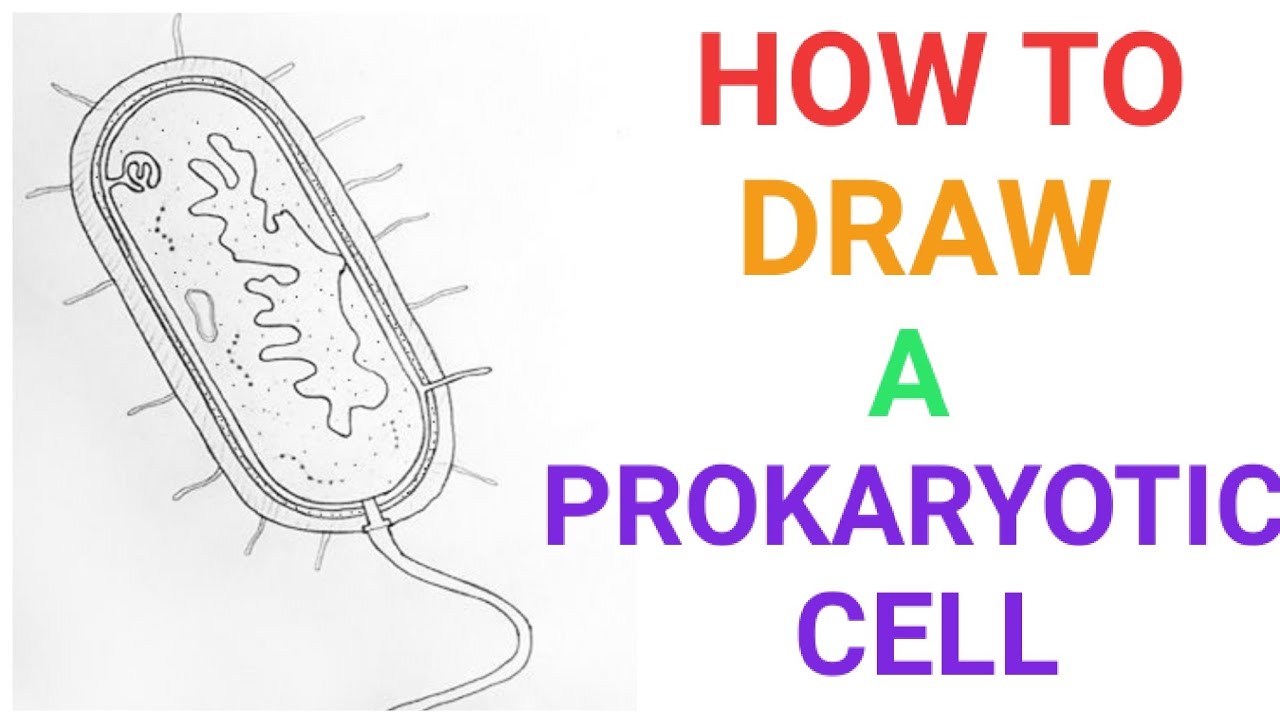
Drawing Of A Prokaryotic Cell - This double layer consists largely of specialized lipids called phospholipids. Prokaryotic cells are not as complex as eukaryotic cells. The other structures shown are present in some, but not all, bacteria. 36k views 3 years ago class 9 diagram. Most prokaryotic cells are much smaller than eukaryotic cells. 175k views 4 years ago diagrams. Web prokaryotic cell structure. Hello friends!!!!in this video, i will be showing you that how to draw a prokaryotic cell very easily.please like, share. Web typical prokaryotic cells range from 0.1 to 5.0 micrometers (μm) in diameter and are significantly smaller than eukaryotic cells, which usually have diameters ranging from 10 to 100 μm. Web i am demonstrating the colorful diagram of prokaryotic cells step by step which you can draw very easily.
Although they are tiny, prokaryotic cells can be distinguished by their shapes. Web both prokaryotic and eukaryotic cells have a plasma membrane, a double layer of lipids that separates the cell interior from the outside environment. Most prokaryotic cells are much smaller than eukaryotic cells. How to draw prokaryotic cell / step by step drawing for beginners. Prokaryotes fall into three basic categories based on their shape, visualized here using scanning electron microscopy: Some archaeal membranes are monolayer rather than bilayer. The diagram of prokaryotic cells helps us to understand the structure of simple prokaryotic organisms and the mechanism by which they interact with the environment. This double layer consists largely of specialized lipids called phospholipids. (a) cocci, or spherical (a pair is shown); Web figure 22.9 common prokaryotic cell types.
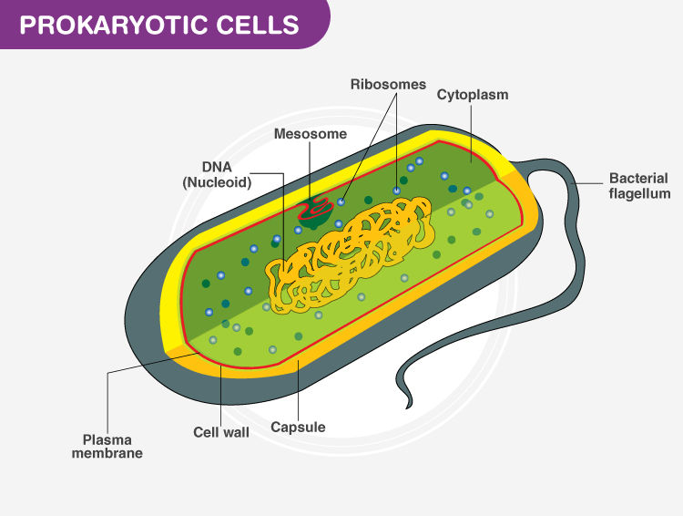
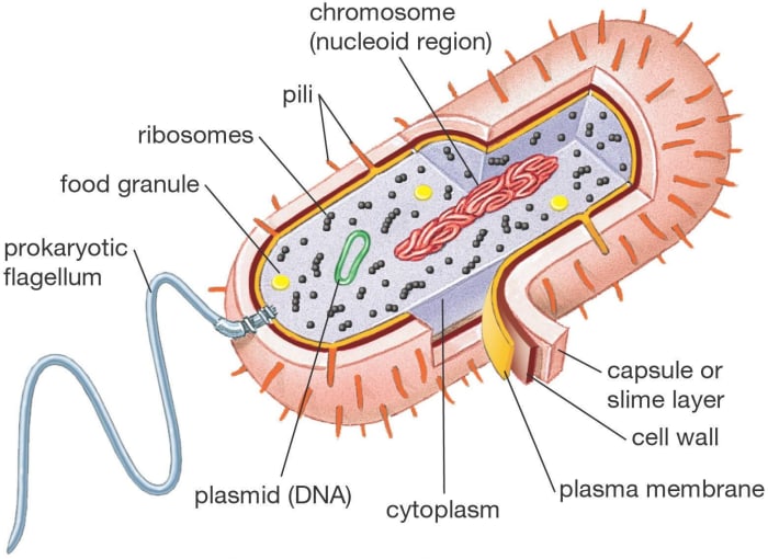
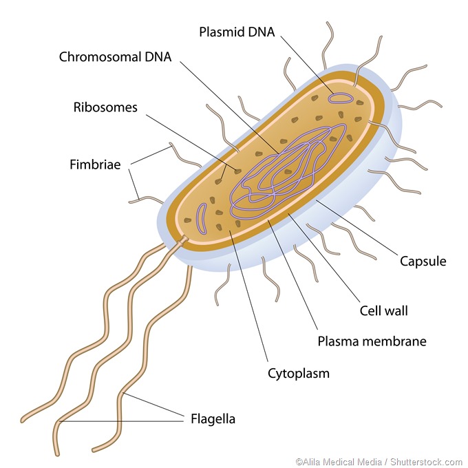
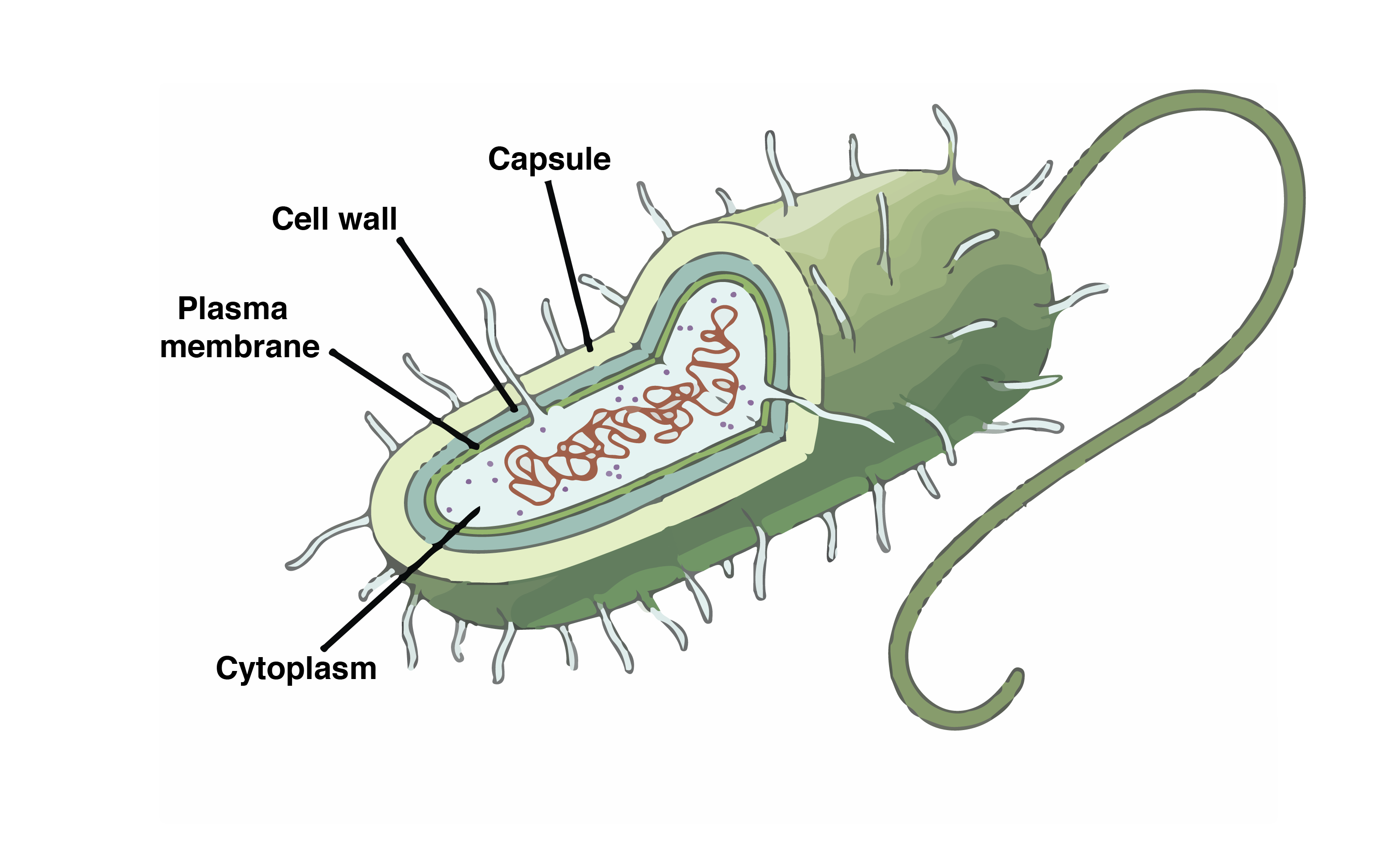
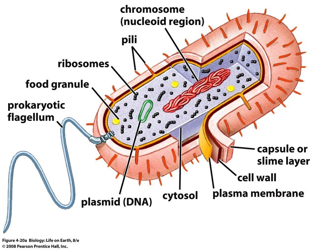
Prokaryotic dna is found in a central part of the cell: The structure called a mesosome was once thought to be an organelle. Web typical prokaryotic cells range from 0.1 to 5.0 micrometers (μm) in diameter and are significantly smaller than eukaryotic cells, which usually have diameters ranging from 10 to 100 μm. This double layer consists largely of specialized lipids called phospholipids. I draw a bacterial cell to show you how to make an accurate biological drawing of a prokaryotic cell. Web figure 4.5 this figure shows the generalized structure of a prokaryotic cell. The other structures shown are present in some, but not all, bacteria. Archaeal membranes have replaced the fatty acids of bacterial membranes with isoprene; The anatomy of a bacterial cell prokaryotic cell structure. All prokaryotes have chromosomal dna localized in a nucleoid, ribosomes, a cell membrane, and a cell wall.
Prokaryotic Cells Definition, Structure, Characteristics, and Examples
Prokaryotic cells are not as complex as eukaryotic cells. In the following sections, we’ll walk through the structure of a prokaryotic cell, starting on the outside and moving towards the inside of the cell. More evidence has convinced most scientists that it is not a true cell structure at all. The other structures shown are present in some, but not.

3.3 Unique Characteristics of Prokaryotic Cells Biology LibreTexts
All prokaryotes have chromosomal dna localized in a nucleoid, ribosomes, a cell membrane, and a cell wall. Most prokaryotic cells are much smaller than eukaryotic cells. How to draw prokaryotic cell step by. Prokaryotes fall into three basic categories based on their shape, visualized here using scanning electron microscopy: Web typical prokaryotic cells range from 0.1 to 5.0 micrometers (μm).
Biology 101 Cells Owlcation Education
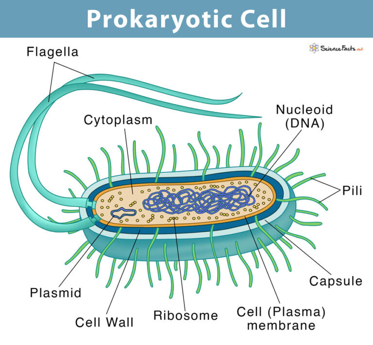
This entry was posted on june 14, 2023 by anne helmenstine (updated on january 23, 2024) the main components of a prokaryotic cell are the plasma membrane, cytoplasm, ribosomes, and nucleoid region. Web prokaryotic cells are distinguished by their shape. The following image is a diagram of a prokaryotic cell; (a) cocci, or spherical (a pair is shown); Some archaeal.
Prokaryotic Cell Definition, Examples, & Structure
Prokaryotes fall into three basic categories based on their shape, visualized here using scanning electron microscopy: Their cell structure is simpler than the cells of animals, plants. This entry was posted on june 14, 2023 by anne helmenstine (updated on january 23, 2024) the main components of a prokaryotic cell are the plasma membrane, cytoplasm, ribosomes, and nucleoid region. Although.
How to draw a prokaryotic cell prokaryotic organism Bacterial cell
This entry was posted on june 14, 2023 by anne helmenstine (updated on january 23, 2024) the main components of a prokaryotic cell are the plasma membrane, cytoplasm, ribosomes, and nucleoid region. Prokaryotes fall into three basic categories based on their shape, visualized here using scanning electron microscopy: Web prokaryotic cell structure. Hello friends!!!!in this video, i will be showing.

Draw A Prokaryotic Cell And Label It
Their cell structure is simpler than the cells of animals, plants. The nucleoid (figure 4.2.1 4.2. How to draw prokaryotic cell step by step for beginners ! The anatomy of a bacterial cell prokaryotic cell structure. Web typical prokaryotic cells range from 0.1 to 5.0 micrometers (μm) in diameter and are significantly smaller than eukaryotic cells, which usually have diameters.
Prokaryotes
And as you can imagine, shape may have something to do with mobility. (a) cocci, or spherical (a pair is shown); Web the prokaryotic cell diagram given below represents a bacterial cell. Bacterial cell anatomy and internal structure. Archaeal membranes have replaced the fatty acids of bacterial membranes with isoprene;
Prokaryotic Gene Structure Chloe's Science
How to draw prokaryotic cell step by. Web the main parts of a prokaryotic cell are shown in this diagram. Their cell structure is simpler than the cells of animals, plants. (a) cocci, or spherical (a pair is shown); It depicts the absence of a true nucleus and the presence of a flagellum that differentiates it from a eukaryotic cell.

Prokaryotic cell structure diagram, Stock vector Colourbox
Prokaryotes fall into three basic categories based on their shape, visualized here using scanning electron microscopy: 355 views 4 months ago isa science. Web prokaryotic cells are distinguished by their shape. Some archaeal membranes are monolayer rather than bilayer. The nucleoid (figure 4.2.1 4.2.
Prokaryotic Cells
These neat, well labelled and colorful diagrams will make your answers look more. This double layer consists largely of specialized lipids called phospholipids. Cells vary regarding other components. Web the main parts of a prokaryotic cell are shown in this diagram. Web figure 22.9 common prokaryotic cell types.
Web The Prokaryotic Cell Diagram Given Below Represents A Bacterial Cell.
(a) cocci, or spherical (a pair is shown); Web many prokaryotic cells have sphere, rod, or spiral shapes (as shown below). We will shortly come to see that this is significantly different in eukaryotes. 355 views 4 months ago isa science.
Their Cell Structure Is Simpler Than The Cells Of Animals, Plants.
In this case, a bacterium. The anatomy of a bacterial cell prokaryotic cell structure. The nucleoid (figure 4.2.1 4.2. Web prokaryotic cells are distinguished by their shape.
Web Prokaryotic Cell Structure.
Web prokaryotic cell diagram to help you remember prokaryotes parts and pieces. I draw a bacterial cell to show you how to make an accurate biological drawing of a prokaryotic cell. It depicts the absence of a true nucleus and the presence of a flagellum that differentiates it from a eukaryotic cell. Web prokaryotic cell diagram.
Web Figure 22.9 Common Prokaryotic Cell Types.
This entry was posted on june 14, 2023 by anne helmenstine (updated on january 23, 2024) the main components of a prokaryotic cell are the plasma membrane, cytoplasm, ribosomes, and nucleoid region. 36k views 3 years ago class 9 diagram. They have no true nucleus as the dna is not contained within a membrane or separated from the rest of the cell, but is coiled up in a region of the cytoplasm called the nucleoid. Modification of work by janice haney carr, dr.