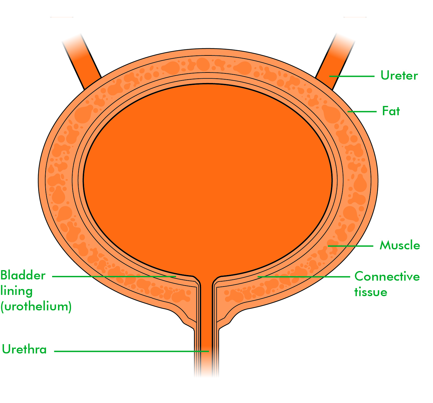
Drawing Of Bladder
Drawing Of Bladder - Reproductive anatomy plays a role in sexual pleasure, getting pregnant, and breastfeeding. It is one of the most elastic organs of the body and is able to increase its volume greatly to accommodate between 600 to 800 ml of urine at maximum capacity. Web drawing of the urinary tract inside the outline of the upper half of a human body. Please provide five to six layers of the cell on the urinary mucosa. Also shown is the prostate. The inside of the left kidney shows the renal pelvis. A set of medical and anatomical parts. The bladder is shown in cross section to reveal interior wall and openings where the ureters empty into the bladder. Urine is made in the renal tubules and collects in the renal pelvis of each kidney. Shows the right and left kidneys, the ureters, the bladder filled with urine, and the urethra passing through the penis.
After the body has taken the food components that it needs, waste products are left behind in the bowel and in the blood. It collects and stores urine from the kidneys before the urine is eliminated through urination. Web browse 1,800+ drawing of bladder stock photos and images available, or start a new search to explore more stock photos and images. In our entire urinary system series,. The urinary system helps rid the body of toxins through urination (peeing). Web the uterus is also shown. Web you can have both types of incontinence. National institute of diabetes and digestive and kidney diseases, national institutes of health. The bladder is part of your urinary. Web urinary bladder (sagittal view) the urinary bladder is a pelvic organ that collects and holds urine before urination.
Please provide five to six layers of the cell on the urinary mucosa. The main parts of the female anatomy can be broken up into outside. The urine flows from the kidneys through the ureters to the bladder. Use the menu to see other pages. Collection of human internal organs hand drawn sketch style. The urinary bladder is a hollow, muscular, and stretchy organ that rests on the pelvic floor. Web first, you should draw the mucosa membrane (folded) of the bladder. Shows the right and left kidneys, the ureters, the bladder filled with urine, and the urethra passing through the penis. Two kidneys are located on either side of the spine near the bottom of the rib cage. It plays two main roles:
Bladder cancer Macmillan's cancer information Blogs Macmillan
As shown in figure \(\pageindex{5}\), urine enters the urinary bladder from the ureters through two ureteral openings on either side of the. You will find drawings of the bladder and its tissue layers. The inside of the left kidney shows the renal pelvis. It plays two main roles: The urinary bladder is a hollow, muscular, and stretchy organ that rests.
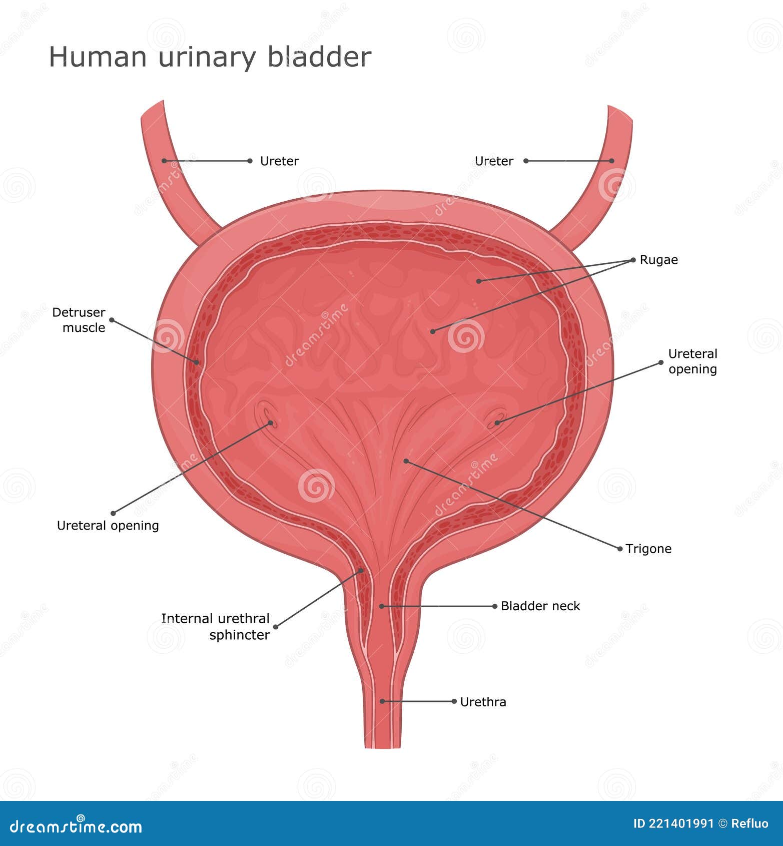
Urinary bladder anatomy stock vector. Illustration of system 221401991
Web urinary bladder (sagittal view) the urinary bladder is a pelvic organ that collects and holds urine before urination. Urine is made in the renal tubules and collects in the renal pelvis of each kidney. Anatomy of the female urinary system showing the kidneys, ureters, bladder, and urethra. It plays two main roles: Let’s draw the lamina propria of the.

Anatomical structure urinary bladder Royalty Free Vector
The urinary bladder is a hollow, muscular, and stretchy organ that rests on the pelvic floor. It can be scary to see blood in your urine, also called hematuria. Web you can have both types of incontinence. It collects and stores urine from the kidneys before the urine is eliminated through urination. In our entire urinary system series,.
Bladder Anatomy Image CustomDesigned Illustrations Creative Market
Web first, you should draw the mucosa membrane (folded) of the bladder. Shows the right and left kidneys, the ureters, the bladder filled with urine, and the urethra passing through the penis. The urinary bladder is a hollow, muscular, and stretchy organ that rests on the pelvic floor. The bladder is shown in cross section to reveal interior wall and.

'Bladder, drawing' Stock Image C002/0616 Science Photo Library
Human internal organs hand drawn sketch vector set. Shows the right and left kidneys, the ureters, the bladder filled with urine, and the urethra passing through the penis. It can be scary to see blood in your urine, also called hematuria. Now, you should try to draw the transitional epithelium on the mucosa of the bladder. The bladder, like the.
What is the Anatomy of the Bladder? (with pictures)
Web female anatomy includes the internal and external structures of the reproductive and urinary systems. Web the bladder is an organ of the urinary system. Web the bladder is shown in cross section to reveal interior wall and openings where the ureters empty into the bladder. It plays two main roles: Transitional epithelium, elastic fibers, and visceral muscle tissue in.

Premium Vector Human bladder hand drawn sketch vector isolated
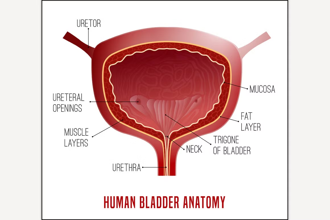
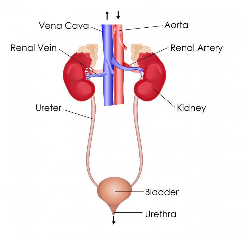
Web the organs of the urinary system include the kidneys, renal pelvis, ureters, bladder and urethra. It collects and stores urine from the kidneys before the urine is eliminated through urination. Let’s draw the lamina propria of the bladder (provide loose connective tissue along with the cells, blood vessels). The kidney and urinary systems help the body to eliminate. Collection.

Human bladder, illustration Stock Image F010/7759 Science Photo
Use the menu to see other pages. Two kidneys are located on either side of the spine near the bottom of the rib cage. The kidneys, ureters, urinary bladder and the urethra.together these organs act to filter blood, remove waste products, create urine and transport urine out from the body. As is the case with most of the pelvic viscera,.

Bladder Anatomy Illustration, Section Stock Vector Illustration of
Web the urinary bladder functions as a storage vessel for urine to delay the frequency of urination. The bladder is shown in cross section to reveal interior wall and openings where the ureters empty into the bladder. Web you can have both types of incontinence. The image shows the urinary tract. In our entire urinary system series,.

Structure of the Bladder
It collects and stores urine from the kidneys before the urine is eliminated through urination. It plays two main roles: The bladder is part of your urinary. Approved by the cancer.net editorial board, 12/2021. It serves as a temporary reservoir for urine produced by the kidneys.when empty, it lies completely within the pelvic cavity, but enlarges upward into the abdominal.
It Serves As A Temporary Reservoir For Urine Produced By The Kidneys.when Empty, It Lies Completely Within The Pelvic Cavity, But Enlarges Upward Into The Abdominal Cavity When Full.it Is The Most Anterior Pelvic Organ,.
The image shows the urinary tract. Now, you should try to draw the transitional epithelium on the mucosa of the bladder. Web the uterus is also shown. Web the organs of the urinary system include the kidneys, renal pelvis, ureters, bladder and urethra.
After The Body Has Taken The Food Components That It Needs, Waste Products Are Left Behind In The Bowel And In The Blood.
The urine flows from the kidneys through the ureters to the bladder. Collection of human internal organs hand drawn sketch style. As shown in figure \(\pageindex{5}\), urine enters the urinary bladder from the ureters through two ureteral openings on either side of the. Web the bladder is shown in cross section to reveal interior wall and openings where the ureters empty into the bladder.
Urine Is Made In The Renal Tubules And Collects In The Renal Pelvis Of Each Kidney.
As is the case with most of the pelvic viscera, there are differences between male and female anatomy of the urinary bladder and urethra. Web the urinary bladder and urethra are pelvic urinary organs whose respective functions are to store and expel urine outside of the body in the act of micturition (urination). Use the menu to see other pages. The urinary system helps rid the body of toxins through urination (peeing).
Approved By The Cancer.net Editorial Board, 12/2021.
Web the bladder is an organ of the urinary system. Web drawing of the urinary tract inside the outline of the upper half of a human body. The bladder is shown in cross section to reveal interior wall and openings where the ureters empty into the bladder. You will find drawings of the bladder and its tissue layers.