Drawing Of Femur Bone
Drawing Of Femur Bone - Web how to draw femur bone step by step | how to draw femur | femur bone👉how to draw diagrams? Web for example, if you’re drawing a femur bone, start with a long oval shape to represent the main body of the bone. The os femoris (thigh bone) articulates proximally with the hip bone (hip joint) and distally with the tibia (knee joint). What does the femur look like? Knee joint icon gray illustration on white. Anterior, medial and posterior view. Web there is a simple way to remember the main anatomical features of the femur using a human stick figure as drawn below. The different parts of the human stick figure correlate with different parts of the femur. Draw 12 more sections, making them smaller as you finish. Let’s walk through the stick figure starting at the head (superior/proximal) and moving to the legs (inferior/distal).
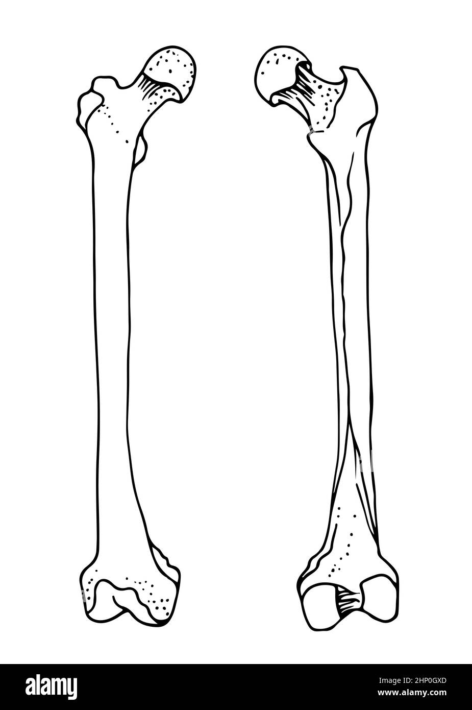
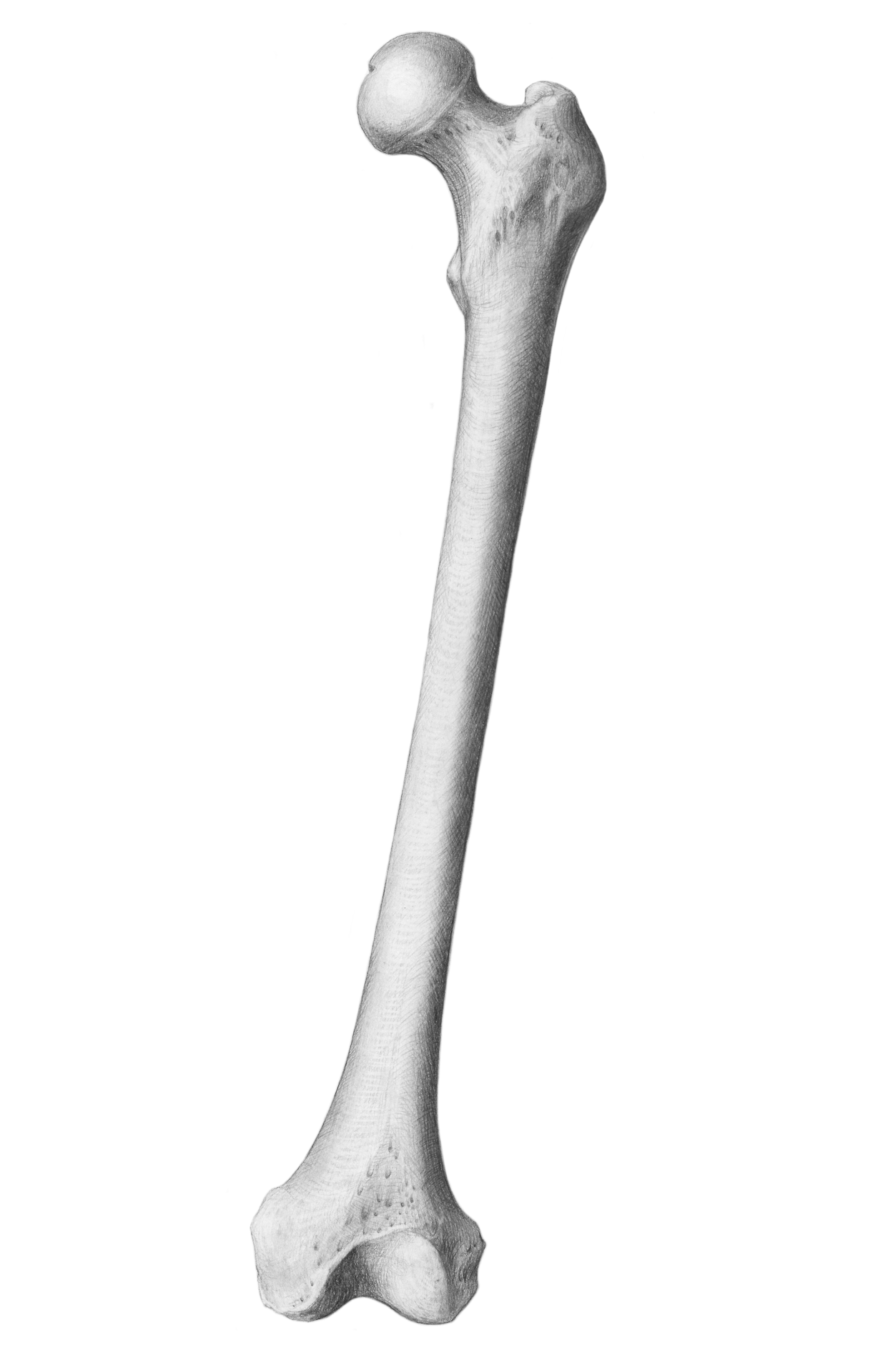
Knee joint icon gray illustration on white. Draw a vertical line and divide it into five parts—these will be the lumbar vertebrae. Where is the femur located? What does the femur look like? It runs from your hip to your knee. Forming the midportion of the femur is a long cylindrical shaft, which arches or curves anteriorly. Draw 12 more sections, making them smaller as you finish. It’s the classic shape used for bones in cartoons: The femur has two rounded ends and a long shaft in the middle. This image shows the right femur from anterior, medial and posterior.
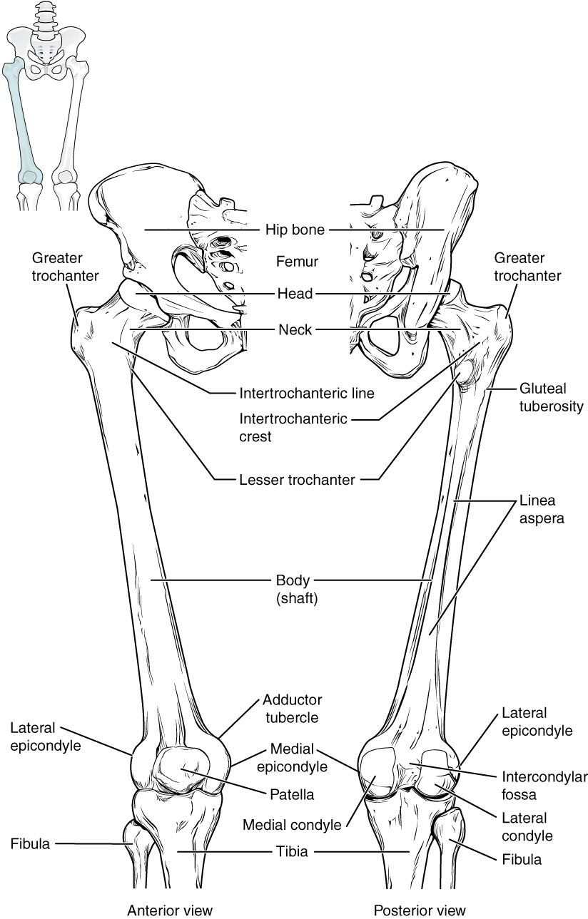
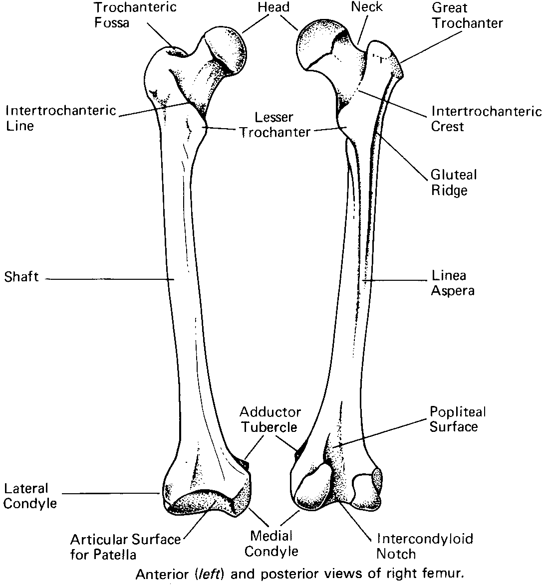
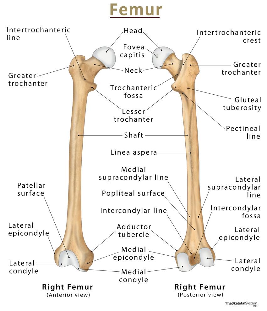
There are also two prominent bony protrusions, the greater and lesser trochanter, that attach to muscles that move the hip and knee. Set of human leg bones isolated on white. Describe the microscopic and gross anatomical structures of bones. Where is the femur located? The different parts of the human stick figure correlate with different parts of the femur. The femur ( os femoris) extends from the hip to the knee and is the longest and strongest bone in the body. The os femoris (thigh bone) articulates proximally with the hip bone (hip joint) and distally with the tibia (knee joint). By the end of this section, you will be able to: The femur is the thigh bone, the largest and strongest bone in the human body. Web learn about the femur bone/thigh bone, what it looks like, where it is located, its definition, functions, parts, & structure, along with labeled pictures
Human femur bones, vector hand drawn illustration isolated on a white
Image and description retrieved from anatomy standard, page femur. There are also two prominent bony protrusions, the greater and lesser trochanter, that attach to muscles that move the hip and knee. Knee joint icon gray illustration on white. Web there is a simple way to remember the main anatomical features of the femur using a human stick figure as drawn.
Bones of the Lower Limb · Anatomy and Physiology
How to draw femur bone human/femour bone human body it is very easy drawing detailed method to help you. Describe the histology of bone tissue, including the function of bone cells and matrix. Identify the gross anatomical features of a bone. Draw a vertical line and divide it into five parts—these will be the lumbar vertebrae. Let’s walk through the.

how to draw femur bone human step by step/femur bone drawing YouTube
It supports the weight of the body and helps you move. Keep your lines light and loose at this stage, as you’ll be refining them later. Web how to draw a skeleton: Once you’re satisfied with the basic shape, start adding more details to your drawing. Web how to draw femur bone step by step | how to draw femur.
Scientific Illustration jennifersmithart Bones Femur, Scapula, and...
Describe the histology of bone tissue, including the function of bone cells and matrix. Outline the discs between the vertebrae. Web how to draw femur bone step by step | how to draw femur | femur bone👉how to draw diagrams? Once you’re satisfied with the basic shape, start adding more details to your drawing. Remember to keep these first lines.

Femur Bone All you will need to know
Knee joint icon gray illustration on white. Forming the midportion of the femur is a long cylindrical shaft, which arches or curves anteriorly. Once you’re satisfied with the basic shape, start adding more details to your drawing. Web there is a simple way to remember the main anatomical features of the femur using a human stick figure as drawn below..

femur bone Femur bone, Basic anatomy and physiology, Anatomy bones
How to draw femur bone human/femour bone human body it is very easy drawing detailed method to help you. Web the femur is the longest, heaviest, and strongest human bone. Antique illustration of human body anatomy bones: The different parts of the human stick figure correlate with different parts of the femur. Knee joint icon gray illustration on white.
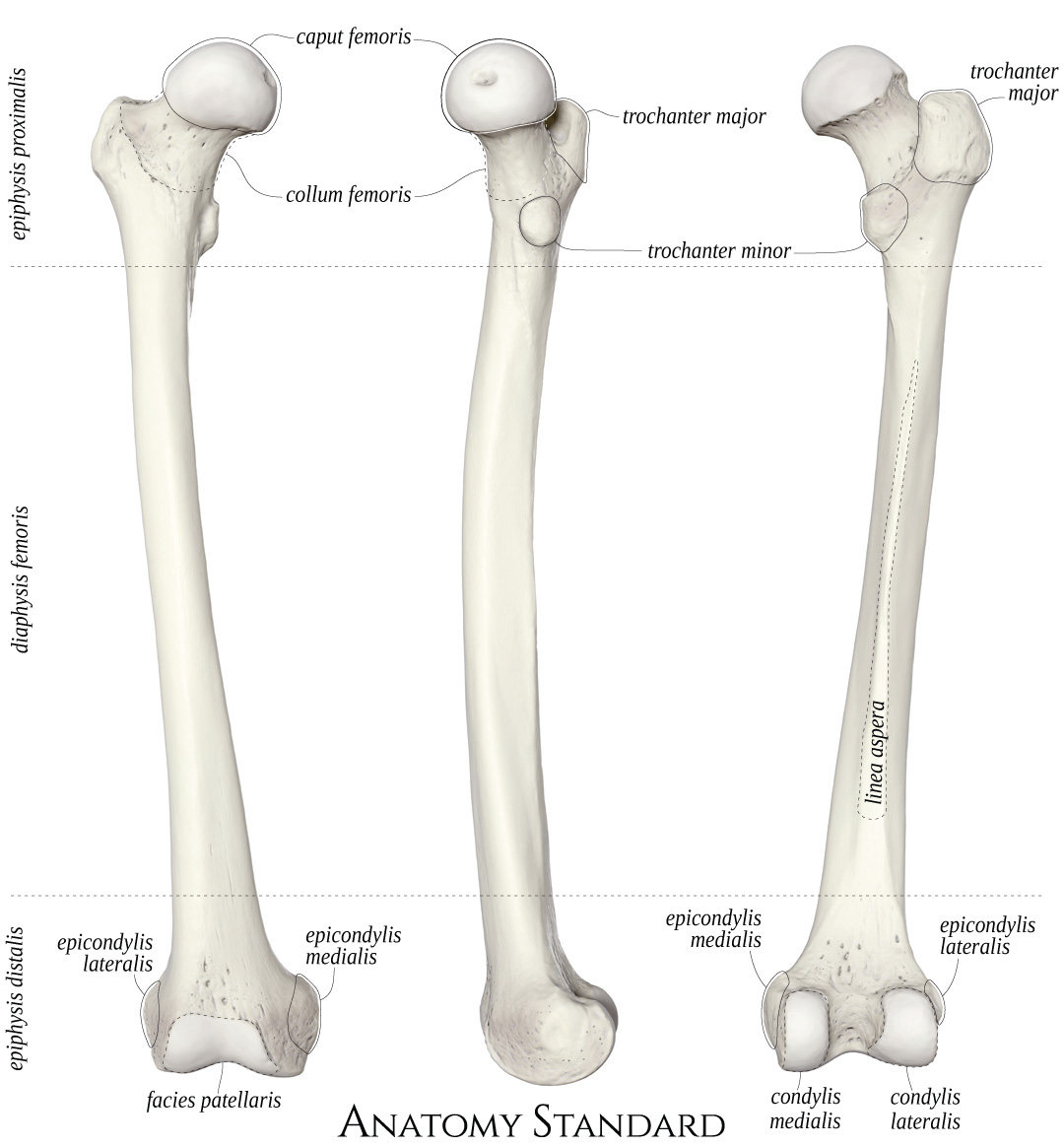
Anatomy Standard Drawing Femur anterior, medial and posterior view
Draw a vertical line and divide it into five parts—these will be the lumbar vertebrae. The femur is the only bone located within the human thigh. Knee joint icon gray illustration on white. Let’s walk through the stick figure starting at the head (superior/proximal) and moving to the legs (inferior/distal). The femur is the only bone in your thigh.
Biology Diagrams,Images,Pictures of Human anatomy and physiology Femur
What does the femur look like? Identify the gross anatomical features of a bone. This image shows the right femur from anterior, medial and posterior. The os femoris (thigh bone) articulates proximally with the hip bone (hip joint) and distally with the tibia (knee joint). Published on 21 june 2023 8 min read.
Radiopaedia Drawing Anatomy of long bones (femur) English labels
This image shows the right femur from anterior, medial and posterior. Draw a vertical line and divide it into five parts—these will be the lumbar vertebrae. Web explore innerbody's 3d anatomical model of the femur bone, the strongest bone in the entire human body. A cylinder with two round bumps at each end. The os femoris (thigh bone) articulates proximally.
Femur Bone Labeled
Compare and contrast compact and spongy bone. Identify the gross anatomical features of a bone. Anterior, medial and posterior view. What does the femur look like? The femur is the only bone located within the human thigh.
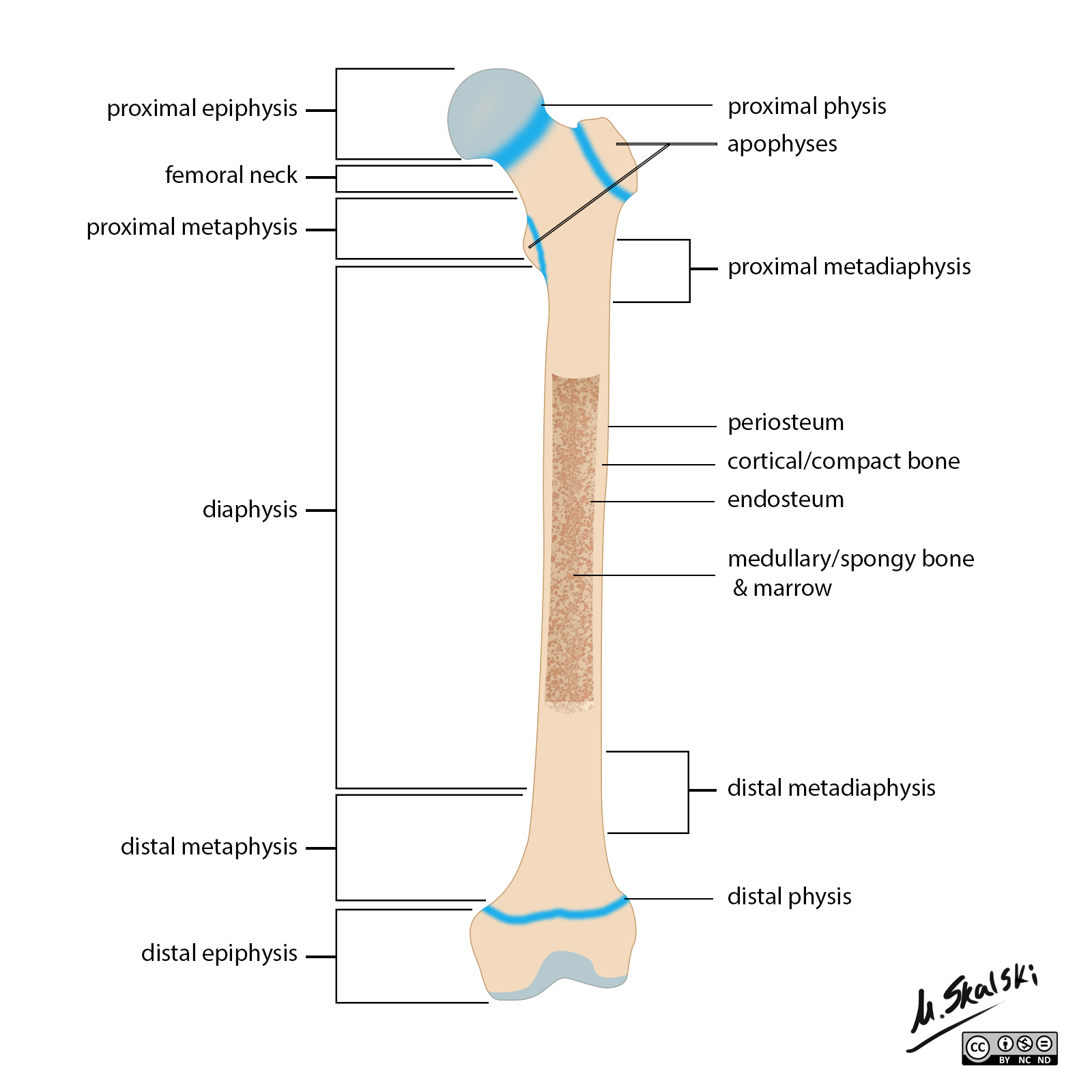
Describe The Microscopic And Gross Anatomical Structures Of Bones.
Published on 21 june 2023 8 min read. It is both the longest and the strongest bone in the human body, extending from the hip to the knee. 21k views 1 year ago #howtodraw #femurbone. Web introduction to femur bone:
There Are Also Two Prominent Bony Protrusions, The Greater And Lesser Trochanter, That Attach To Muscles That Move The Hip And Knee.
Draw 12 more sections, making them smaller as you finish. Knee joint icon gray illustration on white. How to draw femur bone human/femour bone human body it is very easy drawing detailed method to help you. Forming the midportion of the femur is a long cylindrical shaft, which arches or curves anteriorly.
Web How To Draw A Skeleton:
The femur is the only bone located within the human thigh. Web there is a simple way to remember the main anatomical features of the femur using a human stick figure as drawn below. Web learn about the femur bone/thigh bone, what it looks like, where it is located, its definition, functions, parts, & structure, along with labeled pictures Compare and contrast compact and spongy bone.
The Femur Is The Thigh Bone, The Largest And Strongest Bone In The Human Body.
By the end of this section, you will be able to: Knee joint icon gray illustration on white. Set of human leg bones isolated on white. The femur ( os femoris) extends from the hip to the knee and is the longest and strongest bone in the body.