Drawing Of Muscle Cell
Drawing Of Muscle Cell - Myocytes, sometimes called muscle fibers, form the bulk of muscle tissue. Blood vessels and nerves enter the connective tissue and branch in the cell. Web muscle cells, commonly known as myocytes, are the cells that make up muscle tissue. Web the structure of a muscle cell can be explained using a diagram labelling muscle filaments, myofibrils, sarcoplasm, cell nuclei (nuclei is the plural word for the singular nucleus), sarcolemma, and the fascicle of which the muscle fibre is part. Web the muscle cell, or myocyte, develops from myoblasts derived from the mesoderm. Web let's draw a skeletal muscle cell: Thanks for visiting our drawing tutorial in 5 minutes. This article is about skeletal myocytes. Cardiac and skeletal myocytes are sometimes referred to as muscle fibers due to their long and fibrous shape. Web in this video i have shown the simplest way of drawing muscle drawing.
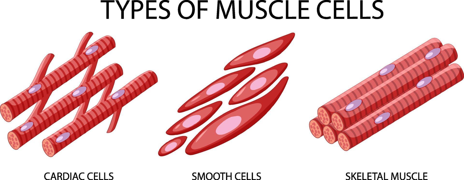
Web how to draw muscle cell step by step. They are bound together by perimysium, a sheath of connective tissue, into bundles called fascicles, which are in. Web however, muscles also enable the heart to beat and can be found in the walls of hollow organs, such as the intestines, uterus and stomach. There are 3 types of muscle cells in the human body; Web relaxing skeletal muscle fibers, and ultimately, the skeletal muscle, begins with the motor neuron, which stops releasing its chemical signal, ach, into the synapse at the nmj. The muscle fiber will repolarize, which closes the gates in. Web each skeletal muscle has three layers of connective tissue (called “mysia”) that enclose it and provide structure to the muscle as a whole, and also compartmentalize the muscle fibers within the muscle (figure 10.3). Muscles work on a macro level, starting with tendons that attach muscles to bones. Excitation signalling of action potentials from the motor neuron are coupled with calcium release. It is the pen diagram of skeletal, smooth and cardiac muscle for class 10, 11 and 12.
These layers cover muscle subunits, individual muscle cells, and myofibrils respectively. These neurons are the site at which the neuron transmits a signal from the brain to the muscle fiber, causing it to contract. This type of cells is found in the wall of internal organs and blood vessels (visceral smooth musculature). Myocytes, sometimes called muscle fibers, form the bulk of muscle tissue. Skeletal muscle tissue is arranged in bundles surrounded by connective tissue. The muscle fiber will repolarize, which closes the gates in. There are 3 types of muscle cells in the human body; The muscular system is responsible for functions such as maintenance of posture, locomotion, and control of various circulatory systems. Web however, muscles also enable the heart to beat and can be found in the walls of hollow organs, such as the intestines, uterus and stomach. Cardiac and skeletal myocytes are sometimes referred to as muscle fibers due to their long and fibrous shape.
Type of muscle cells on white background 5921455 Vector Art at Vecteezy
Web let's draw a skeletal muscle cell: Within muscles, there are layers of connective tissue called the epimysium, perimysium, and endomysium. Web each skeletal muscle has three layers of connective tissue (called “mysia”) that enclose it and provide structure to the muscle as a whole, and also compartmentalize the muscle fibers within the muscle (figure 10.3). Blood vessels and nerves.
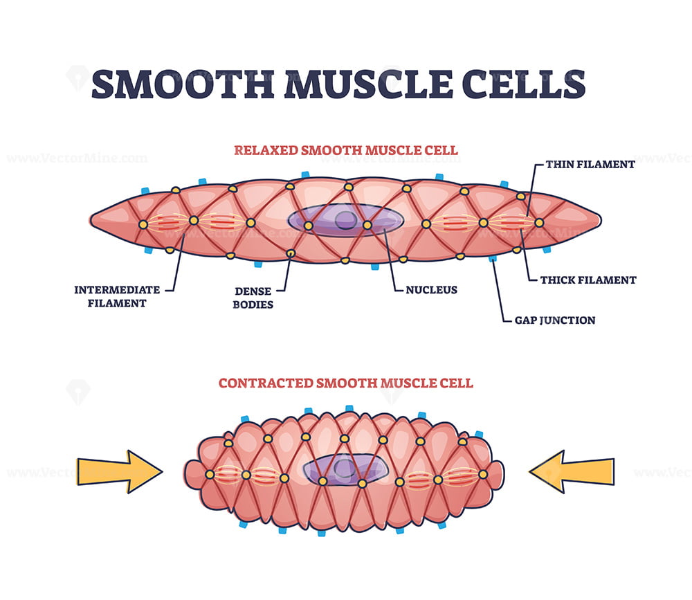
Smooth Muscle Cell Structure
Web the structure of a muscle cell can be explained using a diagram labelling muscle filaments, myofibrils, sarcoplasm, cell nuclei (nuclei is the plural word for the singular nucleus), sarcolemma, and the fascicle of which the muscle fibre is part. Web once the muscle fiber is stimulated by the motor neuron, actin, and myosin protein filaments within the skeletal muscle.
Muscle Cell (Myocyte) Definition, Function & Structure Biology
Web a neuromuscular junction (nmj), also called a myoneural junction, is the connection between a motor neurons and a muscle fibers. 133 views 1 year ago little lectures for 1st semester a&p. They are bound together by perimysium, a sheath of connective tissue, into bundles called fascicles, which are in. Web relaxing skeletal muscle fibers, and ultimately, the skeletal muscle,.

Types of muscle cell diagram 1762350 Vector Art at Vecteezy
They are bound together by perimysium, a sheath of connective tissue, into bundles called fascicles, which are in. The sliding filament theory is the most widely accepted explanation for how this occurs. Web each skeletal muscle has three layers of connective tissue (called “mysia”) that enclose it and provide structure to the muscle as a whole, and also compartmentalize the.

How To Draw Muscle Cell Step by Step YouTube
Web a muscle cell, known technically as a myocyte, is a specialized animal cell which can shorten its length using a series of motor proteins specially arranged within the cell. Web this is what we wanted to get to, but we're going to go even within the muscle cell to see, understand how all the myosin and the actin filaments.

Anatomy Of Muscle Cell The Anatomy Stories
Blood vessels and nerves enter the connective tissue and branch in the cell. Web a muscle cell, also known as a myocyte, is a mature contractile cell in the muscle of an animal. Muscles work on a macro level, starting with tendons that attach muscles to bones. Myocytes, sometimes called muscle fibers, form the bulk of muscle tissue. Web skeletal.

Types of muscle cell diagram 1783902 Vector Art at Vecteezy
The sliding filament theory is the most widely accepted explanation for how this occurs. Myocytes, sometimes called muscle fibers, form the bulk of muscle tissue. Web skeletal muscle fiber structure. They are bound together by perimysium, a sheath of connective tissue, into bundles called fascicles, which are in. What is your request drawing?

Types of muscle cells vector illustration in 2022 Types of muscles
The muscular system is responsible for functions such as maintenance of posture, locomotion, and control of various circulatory systems. Web anatomy of a skeletal muscle cell. Myocytes, sometimes called muscle fibers, form the bulk of muscle tissue. Web this is what we wanted to get to, but we're going to go even within the muscle cell to see, understand how.

Diagram showing types of muscle cells illustration Stock Vector Image
A skeletal muscle cell is long and threadlike with many nuclei and is called a muscle fiber. Web anatomy of a skeletal muscle cell. Like and subscribe our channel to get our latest draw tutorial. Web how to draw muscle cell step by step. There are three layers of connective tissue:

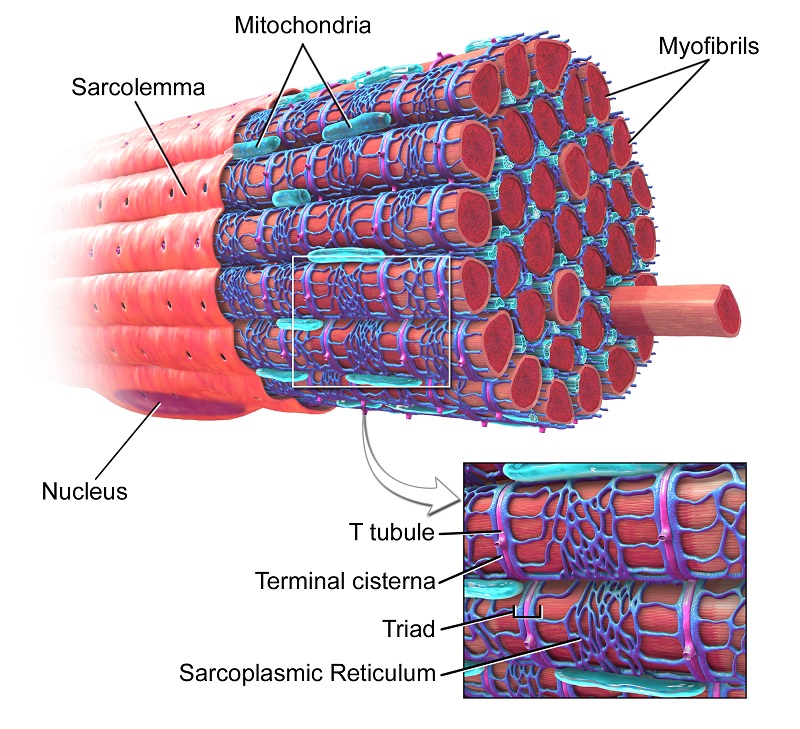
Skeletal Muscle Cell Structure
Web a neuromuscular junction (nmj), also called a myoneural junction, is the connection between a motor neurons and a muscle fibers. Skeletal, smooth, and cardiac (cardiomyocytes). Web skeletal muscle fiber structure. Skeletal muscle tissue is arranged in bundles surrounded by connective tissue. The muscular system is responsible for functions such as maintenance of posture, locomotion, and control of various circulatory.
Web The Structure Of A Muscle Cell Can Be Explained Using A Diagram Labelling Muscle Filaments, Myofibrils, Sarcoplasm, Cell Nuclei (Nuclei Is The Plural Word For The Singular Nucleus), Sarcolemma, And The Fascicle Of Which The Muscle Fibre Is Part.
Web follow our step by step tutorial and be a master draw. Web the muscle cell, or myocyte, develops from myoblasts derived from the mesoderm. Web however, muscles also enable the heart to beat and can be found in the walls of hollow organs, such as the intestines, uterus and stomach. A skeletal muscle cell is long and threadlike with many nuclei and is called a muscle fiber.
Muscles Work On A Macro Level, Starting With Tendons That Attach Muscles To Bones.
Thanks for visiting our drawing tutorial in 5 minutes. Excitation signalling of action potentials from the motor neuron are coupled with calcium release. They are bound together by perimysium, a sheath of connective tissue, into bundles called fascicles, which are in. Web this is what we wanted to get to, but we're going to go even within the muscle cell to see, understand how all the myosin and the actin filaments fit into that muscle cell.
Skeletal Muscle Tissue Is Arranged In Bundles Surrounded By Connective Tissue.
Web anatomy of a skeletal muscle cell. This article is about skeletal myocytes. Web skeletal muscle fiber structure. Web each skeletal muscle has three layers of connective tissue (called “mysia”) that enclose it and provide structure to the muscle as a whole, and also compartmentalize the muscle fibers within the muscle (figure 10.3).
Web Skeletal Muscles Contain Connective Tissue, Blood Vessels, And Nerves.
Within muscles, there are layers of connective tissue called the epimysium, perimysium, and endomysium. These layers cover muscle subunits, individual muscle cells, and myofibrils respectively. The muscle fiber will repolarize, which closes the gates in. In humans and other vertebrates there are three types: