Elodea Cell Drawing
Elodea Cell Drawing - This page is a draft and is under active development. In the circles, you will put your drawings from the microscope. Part b elodea cells. Web login or create a free account. The cell wall, nucleus, and chloroplasts are visible. Draw what you see and label the cell wall and chloroplasts. Web sketch the cell at low and high power. Students compare structures found in each type of cell and create drawings. It is transparent, but you can see where it's pressing the. Web in this lesson, you will observe algal, plant, and animal cells through a microscope.
Record your observations and sketch the following: Web the cell or plasma membrane is present but is not visible because it is thin and in direct contact with the cell wall. Make your drawing 75 mm in its longest dimension. Set up the microscope activity (optional) objectives. Web elodea is a good model to study living plant cells in action. Prepare a wet mount of one leaf from the water plant elodea using the water in which it is kept. Students compare structures found in each type of cell and create drawings. Observe the cells under normal conditions, and make a sketch of what you see. Print out color copies of the large image of elodea cells. Web paper towels or tissues.
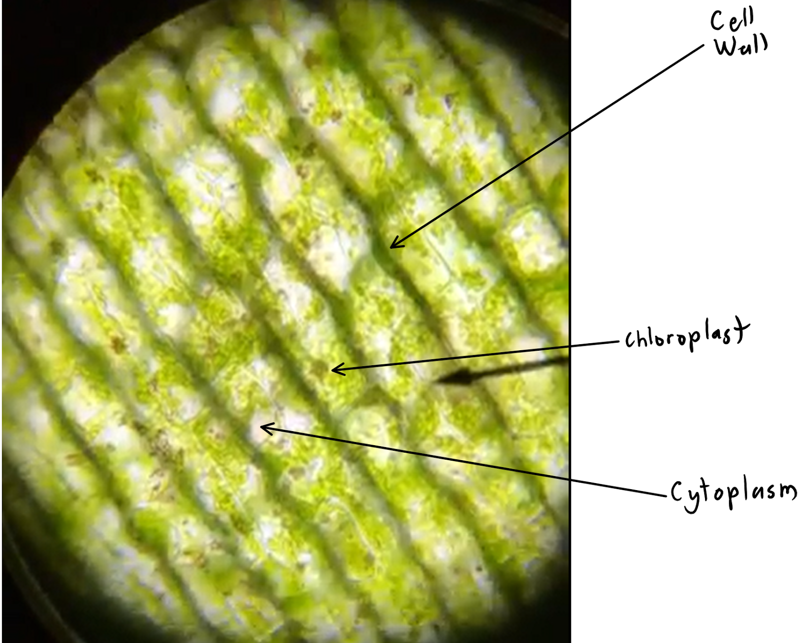
Web elodea leaf cells elodea leaf cells with structures labeled supported by a science education partnership award (sepa) from the national center for research resources, national institutes of health , and the david and lucile packard foundation. This movement is known as cytoplasmic streaming , which distributes nutrients more evenly throughout the. Web login or create a free account. In this demonstration, the aquatic plant called common waterweed, elodea canadensis, serves as a model for cell function. Remove an elodea leaf and place it in the middle of a microscope slide. Label the cell wall, cell membrane, cytoplasm, and chloroplasts in your lab manual. While observing the leaf under the microscope, wick a solution of 6% nacl (sodium chloride) across the slide. Sketch the bacteria at low and high power. Normal elodea cell plasmolyzed elodea (with 6% nacl) Web in this lesson, you will observe algal, plant, and animal cells through a microscope.

Diagram Of Elodea Cell
View a prepared slide of elodea (anacharis), which is an aquarium plant. Elodea is a water plant that grows abundantly in ponds around spokane. Prepare a wet mount of one leaf from the water plant elodea using the water in which it is kept. Set up the microscope activity (optional) objectives. Pick off an entire healthy looking elodea leaf, with.
Plasmolyzed cells of Elodea leaf UWDC UWMadison Libraries
Web elodea leaf cells elodea leaf cells with structures labeled supported by a science education partnership award (sepa) from the national center for research resources, national institutes of health , and the david and lucile packard foundation. Web in this investigation the aquarium water is about 1% salt and 99% water, an elodea cell normally contains 1% salt and 99%.

Elodea leaf cell illustration from a microscope slide. A drop of 10
Be sure to indicate the magnification used and specimen name. Label the cell wall, plasma membrane, cytoplasm, chloroplasts, nucleus (if you see it), central vacuole, and tonoplast. Web elodea leaf cells elodea leaf cells with structures labeled supported by a science education partnership award (sepa) from the national center for research resources, national institutes of health , and the david.

Diagram Of Elodea Cell
Draw your cells to scale. Careful observation should reveal similarities and differences between the cells. To learn about the structure and function of a plant cell and its parts. Set up the microscope activity (optional) objectives. Print and duplicate the student pages.
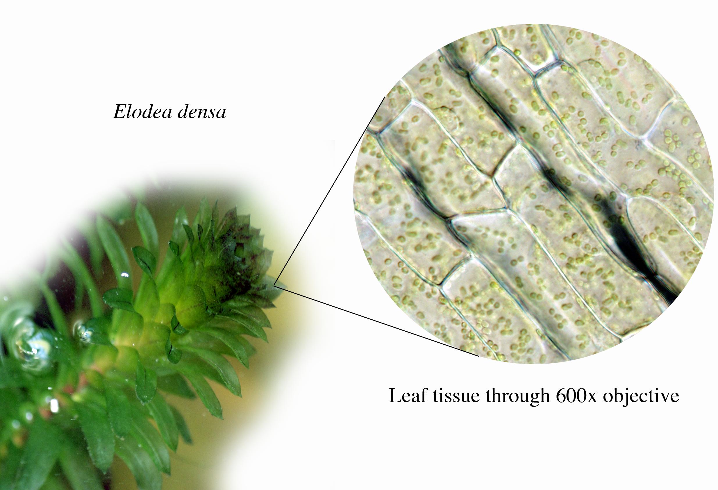
Elodea plant with microscopic view of its leaf cells UWDC UW
Normal elodea cell plasmolyzed elodea (with 6% nacl) Label the cell wall, cell membrane, cytoplasm, and chloroplasts in your lab manual. Web journal of college science teaching—december 2003/january 2004. Set up the microscope activity (optional) objectives. Label the structures in one cell:

Elodea Leaf Cell Under Microscope Labeled Micropedia
Students observe onion cells and elodea cells using the light microscope. Sketch the bacteria at low and high power. Make your drawing 75 mm in its longest dimension. Draw a cell from the azolla in the space below. Label the nucleus, cytoplasm, and cell membrane of a single cell.

Diagram Of Elodea Cell
Students compare structures found in each type of cell and create drawings. Web this elodea leaf cell exemplifies a typical plant cell. Label the nucleus, cytoplasm, and cell membrane of a single cell. Draw your cells to scale. In this lab, you will view cells from elodea, which is a water plant and your cheek cells (animal cells).
![[DIAGRAM] Label Diagram Of Elodea Cells](https://diagramweb.net/img/diagram-of-elodea-cell-5.png)
[DIAGRAM] Label Diagram Of Elodea Cells
A nucleus is present but difficult to see because this preparation is not stained and the chlorophyll masks other structures in the cell. Prepare a wet mount of one leaf from the water plant elodea using the water in which it is kept. Label the structures in one cell: Print and duplicate the student pages. This movement is known as.
![[DIAGRAM] Label Diagram Of Elodea Cells](https://schematron.org/image/elodea-leaf-cell-diagram-12.jpg)
[DIAGRAM] Label Diagram Of Elodea Cells
Normal elodea cell plasmolyzed elodea (with 6% nacl) While observing the leaf under the microscope, wick a solution of 6% nacl (sodium chloride) across the slide. Web paper towels or tissues. Label the cytoplasm and cell wall of a single cell. Students observe onion cells and elodea cells using the light microscope.
Elodea Leaf Cell Diagram Wiring Diagram Pictures
Web login or create a free account. Print and duplicate the student pages. Web plant cell lab. In this experiment, you will see chloroplasts moving in the elodea cells as they begin to photosynthesize. You also will compare the structures of the cells and discuss whether their structures are suited to their functions.
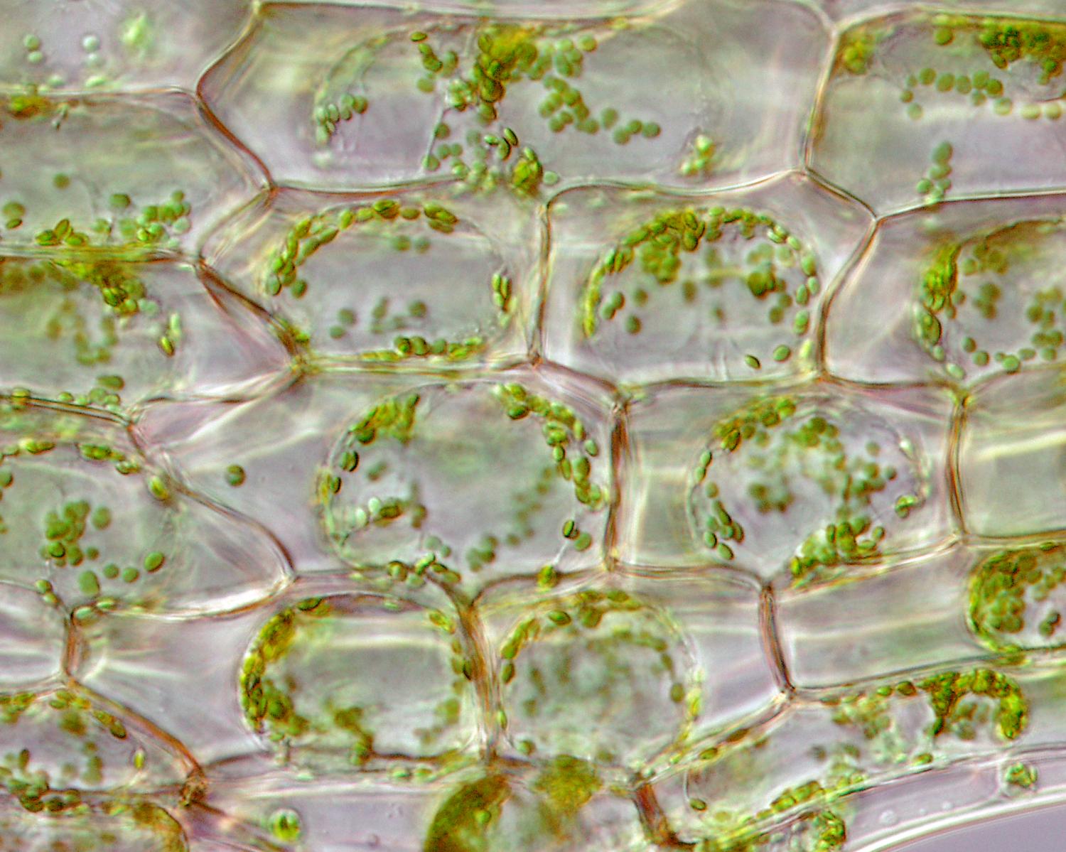
Web Above Is A Cell From The Aquatic Plant Elodea.
The numerous green chloroplasts allow the cell to make its own food (by photosynthesis). Label the structures in one cell: Label the cytoplasm and cell wall of a single cell. Part a onion cells.
Define Cell Membrane, Cell Wall, And Chloroplast.
Draw a cell from the azolla in the space below. Web in this investigation the aquarium water is about 1% salt and 99% water, an elodea cell normally contains 1% salt and 99% water on the inside, the salt water solution is 6% salt and 94% water, and distilled water is 100% water. Using a pipette, drop fresh water on top of the elodea. These are chloroplasts (four are indicated and labeled in the image).
In The Circles, You Will Put Your Drawings From The Microscope.
Part b elodea cells. Set up the microscope activity (optional) objectives. The plant clearly exhibits cell activity by visibly demonstrating cytoplasmic streaming, which occurs in animals, plants, and protista. Print out color copies of the large image of elodea cells.
Prepare A Wet Mount Of One Leaf From The Water Plant Elodea Using The Water In Which It Is Kept.
Web view under the microscope and sketch the cells at each magnification. This image shows cells in the leaf of an aquatic plant, elodea. Draw your cells to scale. Web journal of college science teaching—december 2003/january 2004.