Epithelial Cell Drawing
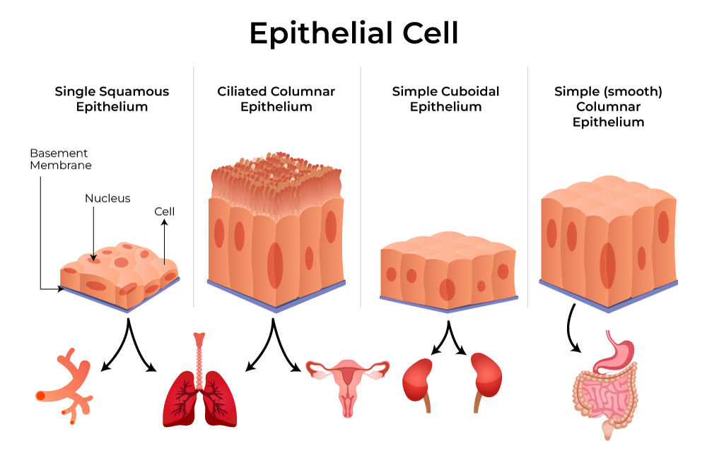
Epithelial Cell Drawing - Cell shapes can be squamous (flattened and thin), cuboidal (boxy, as wide as it is tall), or columnar (rectangular, taller than it is wide). Be able to recognize and classify different epithelial tissues. Web there are three basic shapes used to classify epithelial cells. The cells in this tissue are tightly packed within a thin ecm. A) schematic diagram of eyeball organ culture. Web epithelial cells form from ectoderm, mesoderm, and endoderm, which explains why epithelial line body cavities and cover most body and organ surfaces. The epithelium is a type of body tissue that forms the covering on all internal and external surfaces of your body, lines body cavities and hollow organs and is the major tissue in glands. O describe the structure of microvilli, cilia, and other apical specializations of epithelial cells. O classify morphological types of epithelia based on the number of cell layers, shape of apical cells, and presence of surface specializations. Epithelial cells are often associated with the skin (the epidermis).
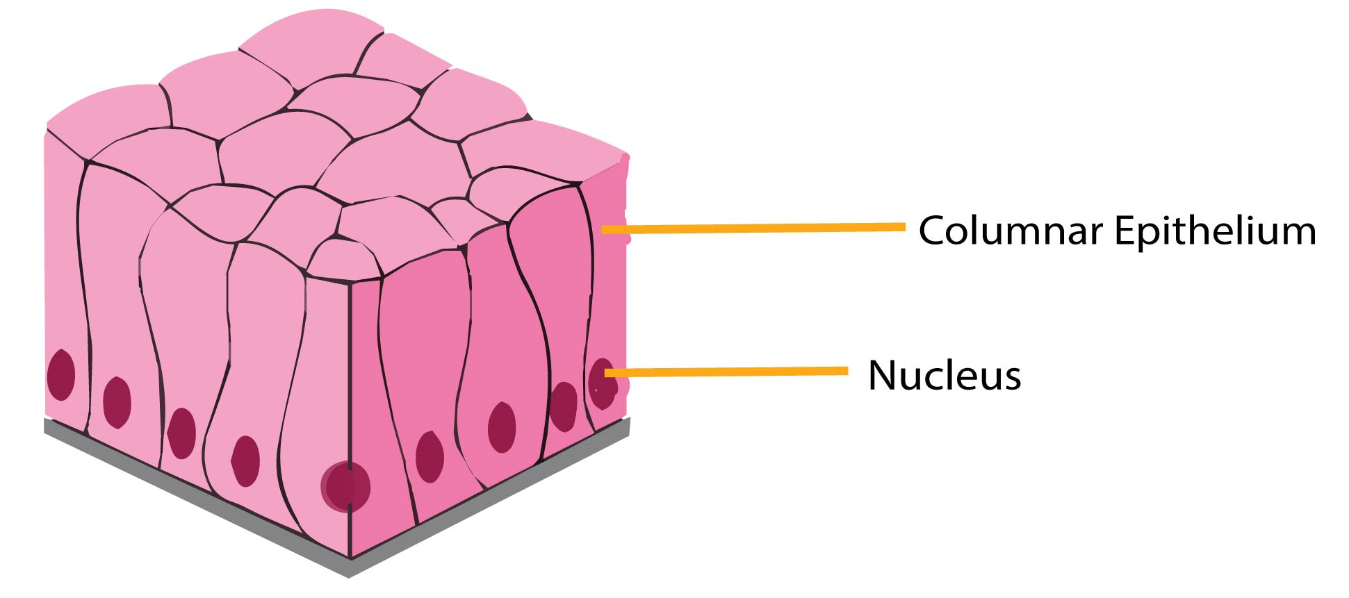
Simple columnar epithelium consists of a single layer of cells that are taller than they are wide, with an oval nucleus usually located towards the basal region of the cell. O classify morphological types of epithelia based on the number of cell layers, shape of apical cells, and presence of surface specializations. O describe the structure of microvilli, cilia, and other apical specializations of epithelial cells. Web the main difference between epithelial and connective tissue is in cells, that they are made up of and their functions. First, epithelial tissue is highly cellular, with little or no extracellular material present between cells. Cell shapes can be squamous (flattened and thin), cuboidal (boxy, as wide as it is tall), or columnar (rectangular, taller than it is wide). Web how to draw simple epithelial tissues. Second, adjoining cells form specialized intercellular connections called cell junctions. Epithelial cells are localised to three distinct areas of the body. 19k views 2 years ago cell biology.
Web epithelium or epithelial tissue is a thin, continuous, protective layer of compactly packed cells with little extracellular matrix. Web how to draw simple epithelial tissues. Web epithelial cells form from ectoderm, mesoderm, and endoderm, which explains why epithelial line body cavities and cover most body and organ surfaces. Lining interior tracts which open to the exterior, such as the respiratory, gastrointestinal and genitourinary tracts. Web corneal permeation and ocular tolerance in vivo. A cuboidal epithelial cell looks close to a square. Epithelial cells are localised to three distinct areas of the body. Epithelial cells are squamous, cuboidal, or columnar in shape and form either single or multiple layers. Distinguish between tight junctions, anchoring junctions, and gap junctions. A) schematic diagram of eyeball organ culture.

epithelial tissue, drawing Stock Image C015/2525 Science Photo
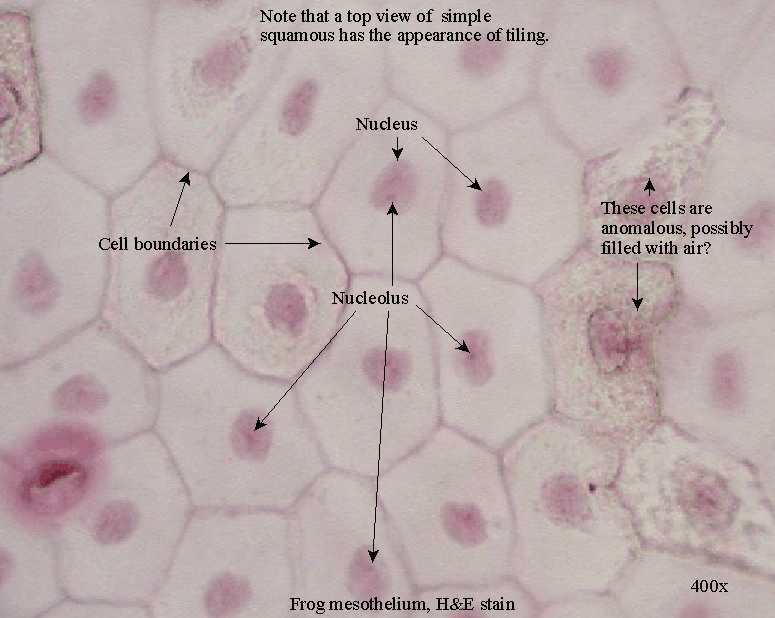
Cells in connective tissue are immersed in an indefinite matrix in addition to cartilaginous, collagen, elastic or fatty tissues. A squamous epithelial cell looks flat under a microscope. They cover organs and body cavities. O classify morphological types of epithelia based on the number of cell layers, shape of apical cells, and presence of surface specializations. Web most epithelial tissues.
![[DIAGRAM] Microscope Epithelial Cell Diagram](https://www.onlinebiologynotes.com/wp-content/uploads/2018/02/Epithelium.jpg)
[DIAGRAM] Microscope Epithelial Cell Diagram
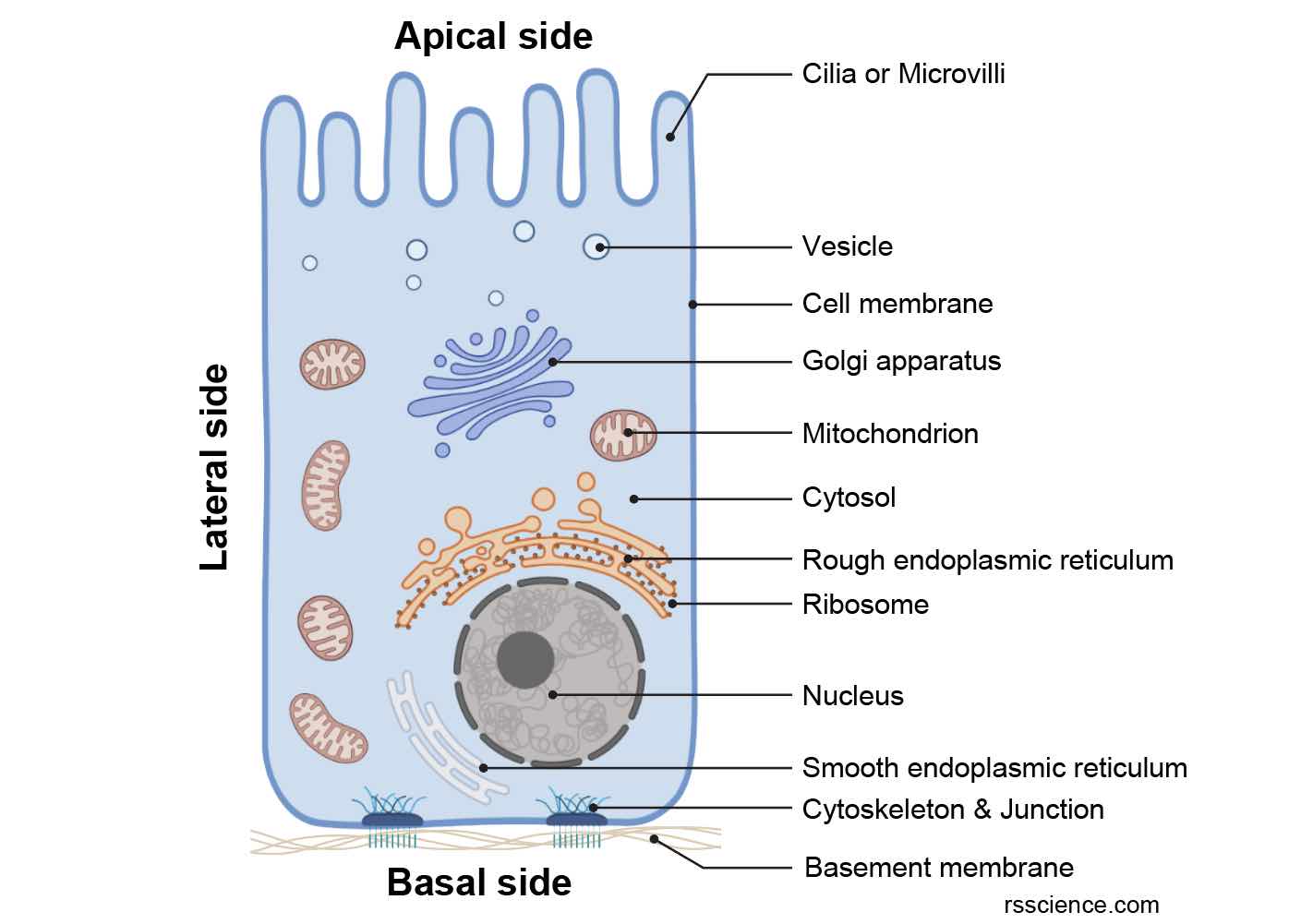
Web the main difference between epithelial and connective tissue is in cells, that they are made up of and their functions. Particular structures found in some epithelial cells are an adaptation to specific functions. Explain the structure and function of epithelial tissue. These cells form continuous layers called epithelia that cover the body’s surfaces, line cavities, and form glands. [1].
Epithelial Tissue Structure, Types and Function (With Diagram) Free
Web how to draw simple epithelial tissues. Covering the whole external surface of the body, as part of the skin. Web most epithelial tissues are essentially large sheets of cells covering all the surfaces of the body exposed to the outside world and lining the outside of organs. In addition, they are in glands. Web corneal permeation and ocular tolerance.
Epithelium What It Is, Function & Types
Epithelial cells are localised to three distinct areas of the body. Web corneal permeation and ocular tolerance in vivo. Epithelial cells line the surfaces of organs in the body and function as a protective barrier. Cells in connective tissue are immersed in an indefinite matrix in addition to cartilaginous, collagen, elastic or fatty tissues. Epithelial cells are squamous, cuboidal, or.
Epithelial Tissue Characteristics, Types, and Functions Owlcation
[1] there are many arrangements of epithelial cells, such as squamous, cuboidal, and columnar, that organize as simple, stratified, pseudostratified, and transitional. However, the epidermis is only one of the many types of epithelial tissue. O explain the composition and function of the basement membrane. A squamous epithelial cell looks flat under a microscope. Web the main difference between epithelial.
Epithelial Tissues Simple Tissue Biology Tissue Types Anatomy And
Web there are three basic shapes used to classify epithelial cells. Epithelial cells are squamous, cuboidal, or columnar in shape and form either single or multiple layers. Epithelial cells are localised to three distinct areas of the body. Web regardless of its location and function, all epithelial tissue shares important structural features. Create professional science figures in minutes with biorender.
Describe various types of epithelial tissues with the help of labeled
19k views 2 years ago cell biology. Be able to correlate different types of epithelia with their locations and functions. They function in protection, secretion, absorption, and transportation of substances. Web most epithelial tissues are essentially large sheets of cells covering all the surfaces of the body exposed to the outside world and lining the outside of organs. Create professional.
Epithelial Tissue Introduction, Characteristics, Types, Importance
Epithelial cells are often associated with the skin (the epidermis). O classify morphological types of epithelia based on the number of cell layers, shape of apical cells, and presence of surface specializations. A squamous epithelial cell looks flat under a microscope. A cuboidal epithelial cell looks close to a square. Epithelial cells are localised to three distinct areas of the.
Epithelium Definition, Characteristics, Cell Structures, Types, and
There are several different types of epithelial cells based on their shape and arrangement. Epithelial cells have many roles in an organism, such as playing a part in secretion, absorption, sensation, protection and. [1] there are many arrangements of epithelial cells, such as squamous, cuboidal, and columnar, that organize as simple, stratified, pseudostratified, and transitional. Like all types, it is.
Epithelial Tissue Anatomy & Physiology
They cover organs and body cavities. However, the epidermis is only one of the many types of epithelial tissue. Web how to draw simple epithelial tissues. Be able to recognize and classify different epithelial tissues. This plasticity is crucial for embryonic development and wound healing, but also underlies the acquisition of several malignant.
They Cover Organs And Body Cavities.
They function in protection, secretion, absorption, and transportation of substances. Distinguish between simple epithelia and stratified epithelia, as well as between squamous, cuboidal, and columnar epithelia. Web there are three basic shapes used to classify epithelial cells. A cuboidal epithelial cell looks close to a square.
Epithelial Cells Have Many Roles In An Organism, Such As Playing A Part In Secretion, Absorption, Sensation, Protection And.
A squamous epithelial cell looks flat under a microscope. Web epithelial cells form from ectoderm, mesoderm, and endoderm, which explains why epithelial line body cavities and cover most body and organ surfaces. O describe the structure of microvilli, cilia, and other apical specializations of epithelial cells. Epithelial cells are localised to three distinct areas of the body.
A Columnar Epithelial Cell Looks Like A Column Or A Tall Rectangle.
Web most epithelial tissues are essentially large sheets of cells covering all the surfaces of the body exposed to the outside world and lining the outside of organs. A) schematic diagram of eyeball organ culture. Explain the structure and function of epithelial tissue. Epithelial cells line the surfaces of organs in the body and function as a protective barrier.
Epithelial Cells Are Squamous, Cuboidal, Or Columnar In Shape And Form Either Single Or Multiple Layers.
Web simple epithelium has only one cell layer where every cell is in direct contact with the underlying basement membrane. First, epithelial tissue is highly cellular, with little or no extracellular material present between cells. Web epithelial cells create the covering layer for your body surfaces. Be able to correlate different types of epithelia with their locations and functions.