Esophagus Drawing
Esophagus Drawing - It functions as part of your digestive system. Select from premium esophagus drawing images of the highest quality. When the patient is upright, the esophagus is usually between 25 to 30. Abdominal which travels past the diaphragm into the abdomen, reaching the stomach. Identification points of esophagus slide. Browse 66 esophagus drawing photos and images available, or start a new search to explore more photos and images. March 30, 2024 fact checked. Tunica serosa or adventitia of esophagus. In an adult, the esophagus is usually around 25 to 30 centimeters in length and can measure up to about 2 centimeters in width. Web the esophagus is a long, thin, and muscular tube that connects the pharynx (throat) to the stomach.
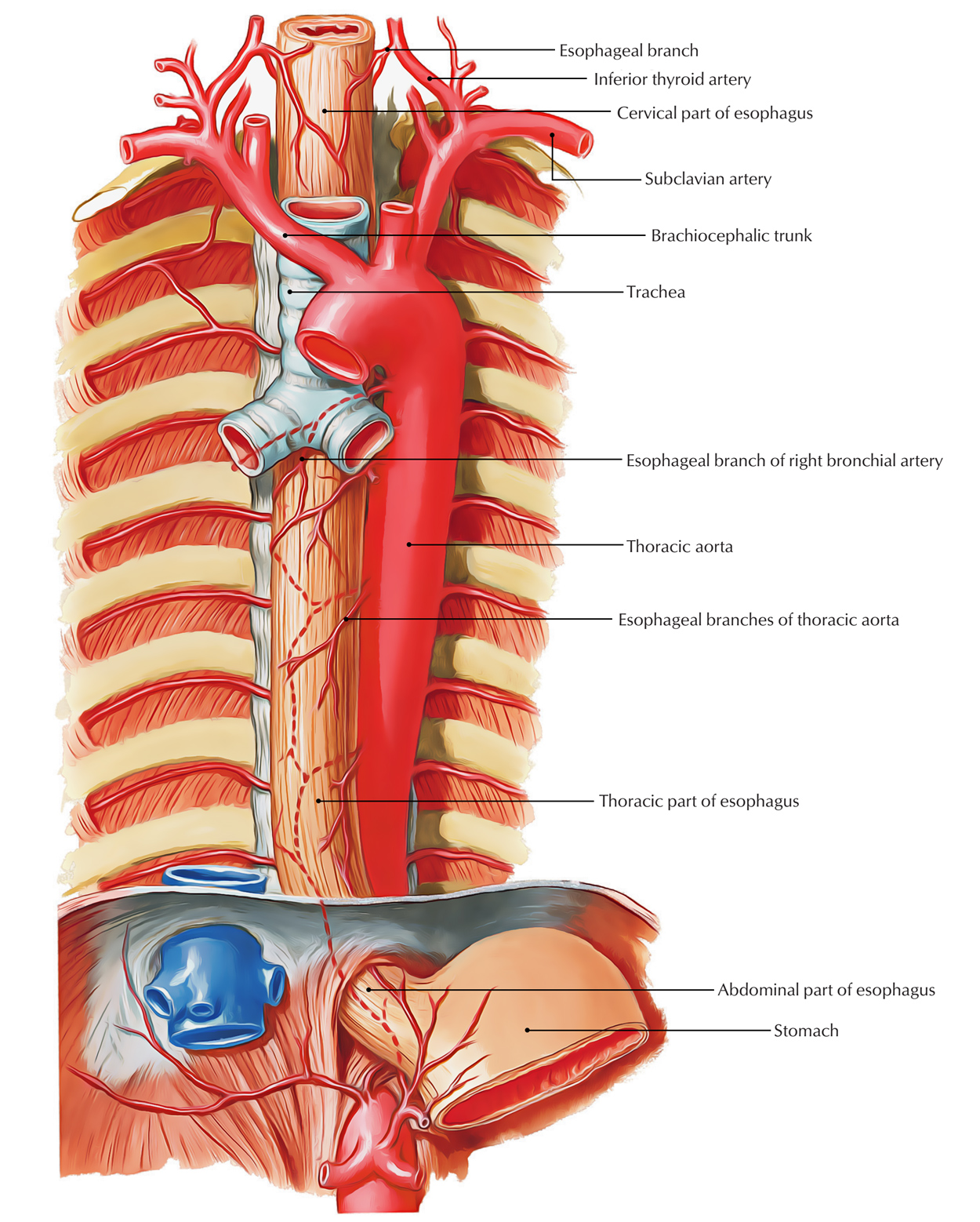
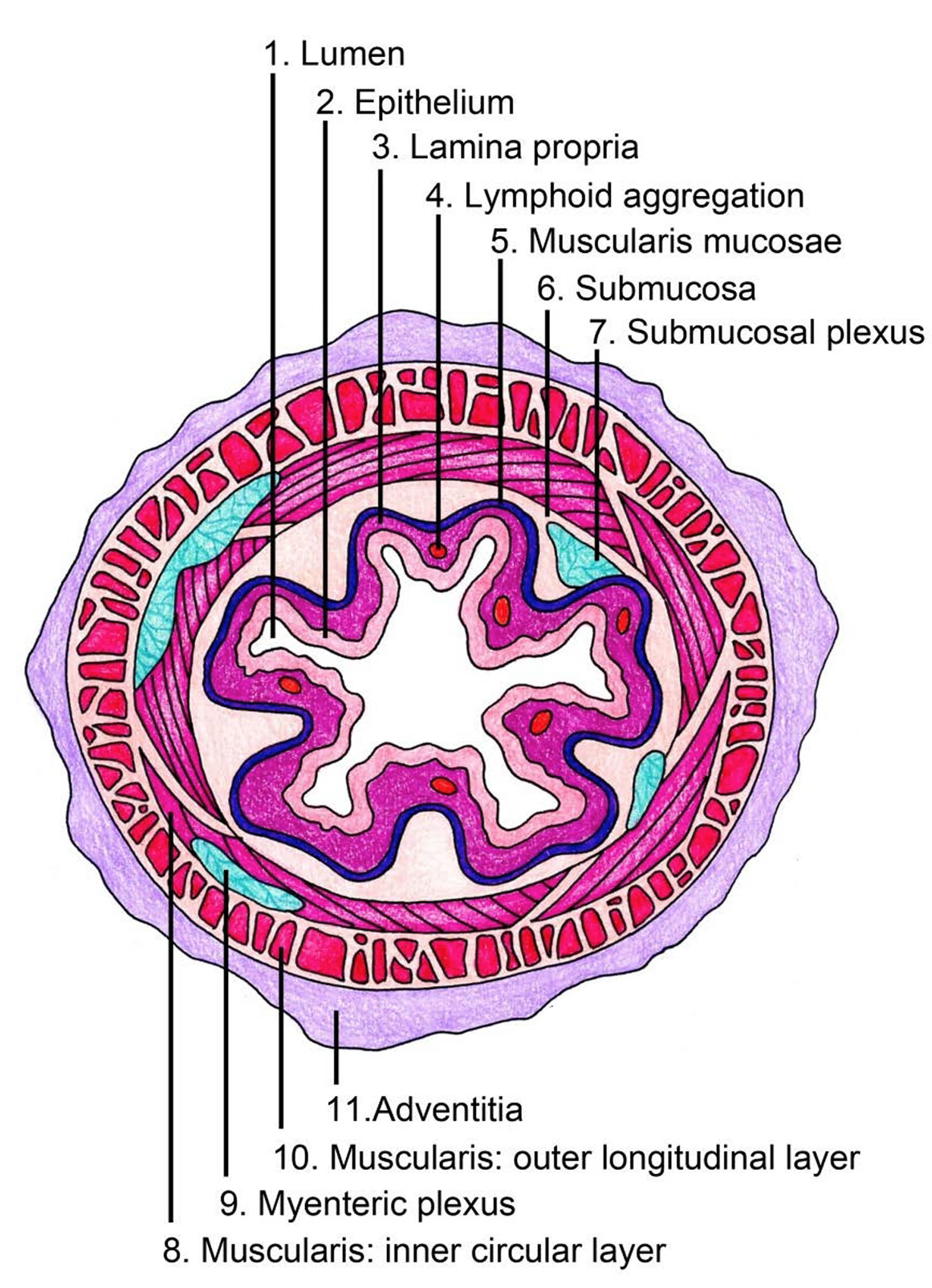
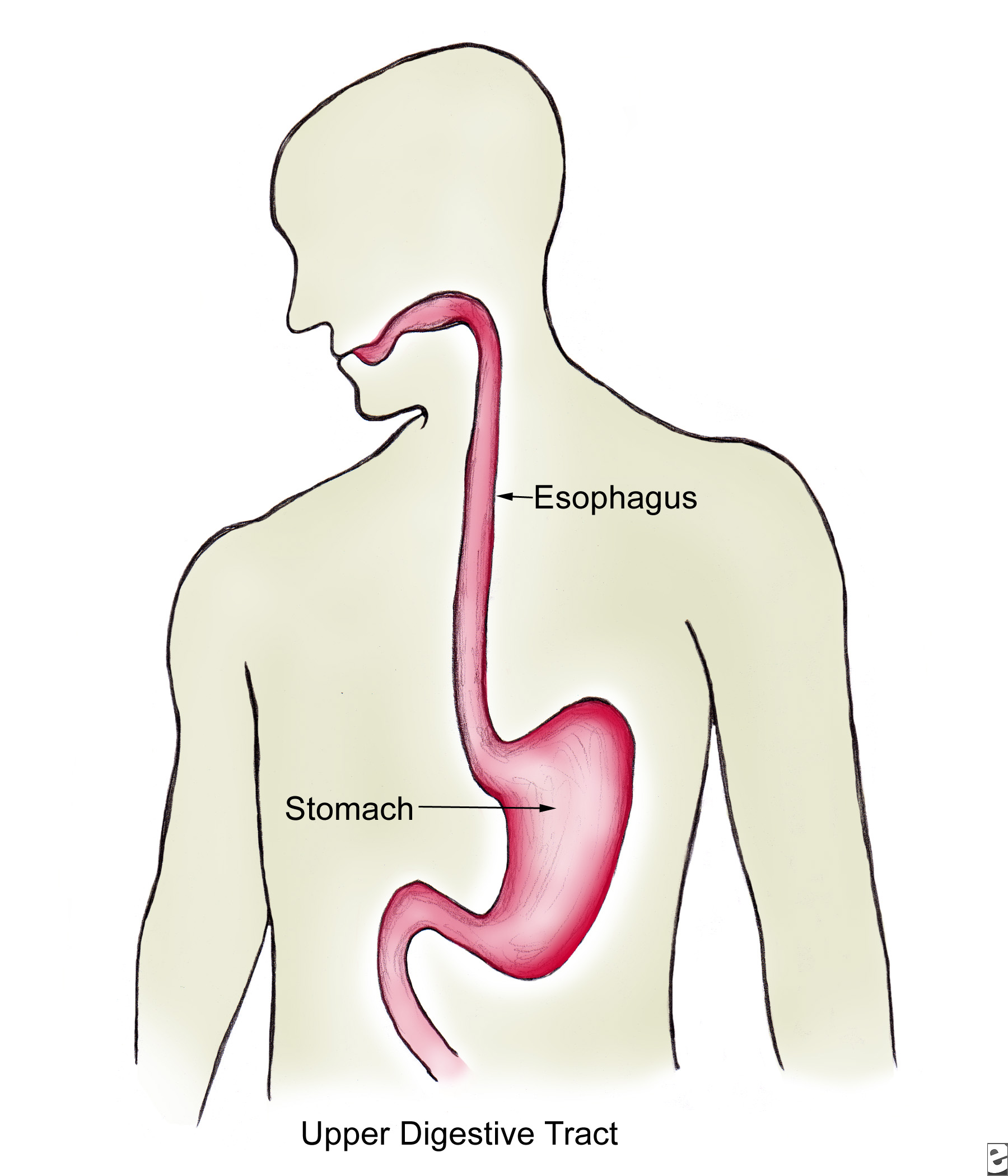
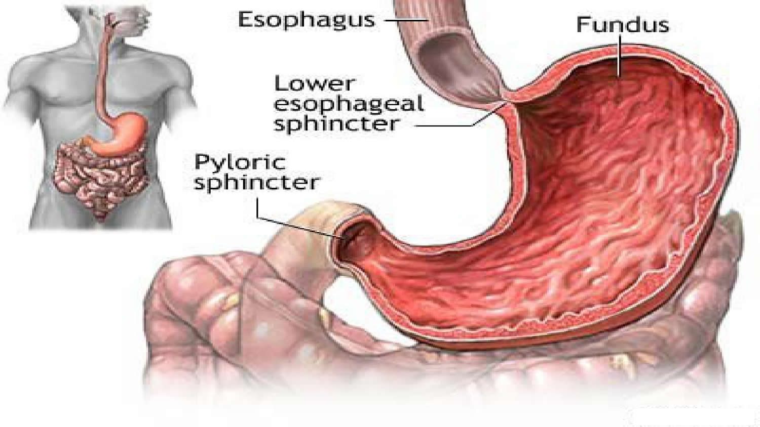
The esophagus is the muscular tube that connects the back of the throat (or pharynx) with the stomach. Tunica muscularis layer of esophagus slide. Drawing shows the pharynx (throat), esophagus, and stomach. Its main job is to deliver food, liquids, and saliva to the rest of the digestive system. The esophagus is a muscular tube about ten inches (25 cm.) long, extending from the hypopharynx to the stomach. Upper and lower human gastrointestinal tract. Web the esophagus is a long, thin, and muscular tube that connects the pharynx (throat) to the stomach. Web table of contents. The esophagus lies posterior to the trachea and the heart and passes through the mediastinum and the hiatus, an opening in the diaphragm, in its descent from the thoracic to the abdominal cavity. Layers of esophagus histology with labeled diagram.
Select from premium esophagus drawing images of the highest quality. Esophagus drawing stock photos are available in a variety of sizes and formats to fit your needs. Its main job is to deliver food, liquids, and saliva to the rest of the digestive system. Thoracic which is located in the thorax, more specifically in the mediastinum. Web anatomy of the esophagus; Base of the tongue, superior view. In an adult, the esophagus is usually around 25 to 30 centimeters in length and can measure up to about 2 centimeters in width. Web median section through the nasal, oral, pharynx and larynx cavities. The esophagus can also widen on its own to allow solids to pass through more easily. The esophagus is muscular, pink in color, and approximately 8 inches long.
Esophagus Earth's Lab
The inner lining of the esophagus is called the mucosa. March 30, 2024 fact checked. (o)esophagi or (o)esophaguses ), colloquially known also as the food pipe, food tube, or gullet, is an organ in vertebrates through which food passes, aided. Base of the tongue, superior view. Browse 2,000 esophagus anatomy photos and images available, or start a new search to.

E.3. Esophagus
Find esophagus drawing stock illustrations from getty images. Cross section of pharynx, esophagus, and larynx. Base of the tongue, superior view. Select from premium esophagus drawing images of the highest quality. Posterior view of pharyngeal muscles.
The Human Esophagus Functions and Anatomy and Problems
Base of the tongue, superior view. (o)esophagi or (o)esophaguses ), colloquially known also as the food pipe, food tube, or gullet, is an organ in vertebrates through which food passes, aided. Layers of esophagus histology with labeled diagram. The esophagus is the muscular tube that connects the back of the throat (or pharynx) with the stomach. The esophagus is divided.
The esophagus Structure of the esophagus
Browse 66 esophagus drawing photos and images available, or start a new search to explore more photos and images. The esophagus ( american english) or oesophagus ( british english, see spelling differences; Location of the esophagus in the human body. (o)esophagi or (o)esophaguses ), colloquially known also as the food pipe, food tube, or gullet, is an organ in vertebrates.
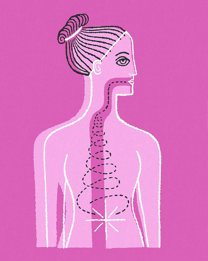
Diagram of Esophagus Drawing by CSA Images Pixels
A pullout shows the mucosa layer, thin muscle layer, submucosa layer, thick muscle layer, and connective tissue layer of the esophagus wall. Posterior view of pharyngeal muscles. Upper and lower human gastrointestinal tract. A medical anatomy diagram of a woman showing the human digestive system. March 30, 2024 fact checked.

esophagus anatomy 2 by speedboy201 on DeviantArt
Drawing shows the pharynx (throat), esophagus, and stomach. Tunica serosa or adventitia of esophagus. A pullout shows the mucosa layer, thin muscle layer, submucosa layer, thick muscle layer, and connective tissue layer of the esophagus wall. Human digestive system woman anatomy diagram. Find esophagus drawing stock illustrations from getty images.

How to Draw Esophagus and Mouth Anotamy Drawing YouTube
It forms an important piece of the gastrointestinal tract and functions as the conduit for food and liquids that have been swallowed into the. Web the esophagus is a hollow muscular tube that transports saliva, liquids, and foods from the mouth to the stomach. (o)esophagi or (o)esophaguses ), colloquially known also as the food pipe, food tube, or gullet, is.

The Mouth, Pharynx, and Esophagus Biology of Aging
Cervical which travels through the neck. Upper and lower human gastrointestinal tract. Abdominal which travels past the diaphragm into the abdomen, reaching the stomach. Web anatomy of the esophagus. The oesophagus or gullet is a muscular canal, about 23 to 25 cm.

Human anatomy esophagus side view on a white Vector Image
Browse 2,000 esophagus anatomy photos and images available, or start a new search to explore more photos and images. The esophagus is a muscular tube about ten inches (25 cm.) long, extending from the hypopharynx to the stomach. Abdominal which travels past the diaphragm into the abdomen, reaching the stomach. Browse 66 esophagus drawing photos and images available, or start.
The Human Esophagus Functions and Anatomy and Problems
The esophagus lies posterior to the trachea and the heart and passes through the mediastinum and the hiatus, an opening in the diaphragm, in its descent from the thoracic to the abdominal cavity. Tunica muscularis layer of esophagus slide. Web the esophagus is a hollow muscular tube that transports saliva, liquids, and foods from the mouth to the stomach. Layers.
The Esophagus Is The Tube That Connects The Mouth And Throat (Pharynx) To The Stomach.
Select from premium esophagus drawing images of the highest quality. The esophagus is divided into three parts: Tunica muscularis layer of esophagus slide. Web table of contents.
Select From Premium Esophagus Drawing Images Of The Highest Quality.
The esophagus is a muscular tube about ten inches (25 cm.) long, extending from the hypopharynx to the stomach. Posterior view of pharyngeal muscles. The esophagus is muscular, pink in color, and approximately 8 inches long. Web the esophagus is a hollow muscular tube that transports saliva, liquids, and foods from the mouth to the stomach.
Thoracic Which Is Located In The Thorax, More Specifically In The Mediastinum.
Drawing shows the pharynx (throat), esophagus, and stomach. Long, extending from the pharynx to the stomach. Web anatomy of the esophagus. Location of the esophagus in the human body.
March 30, 2024 Fact Checked.
Cervical which travels through the neck. The esophagus lies posterior to the trachea and the heart and passes through the mediastinum and the hiatus, an opening in the diaphragm, in its descent from the thoracic to the abdominal cavity. Identification points of esophagus slide. It forms an important piece of the gastrointestinal tract and functions as the conduit for food and liquids that have been swallowed into the.