Eyeball Anatomy Drawing
Eyeball Anatomy Drawing - Pick the right drawing tools. Web © 2023 google llc. The first thing we're going to draw is the white part of the eye, which is known as the sclera. Science & technology 3d models. #ilustrator #art #artist #arte #drawingoftheday #anime #animeart #draw #drawing #manga #instaart #digitalart.. A hole in the middle of the iris that changes size to let in more or less light. Web discovering the underlying anatomy of the eyeball can help you bring life and feeling into your drawings, from the corners of the eye to the pupils. Start with a basic sketch. Hi friends,in this video i will show you how to draw a eye diagram very easy way, its a step by step tutorial to draw diagram of eye anatomy. Web in this video, we're going to talk about the structure of the eye.
Science & technology 3d models. Refractive media of the eyeball. Drawing digitally with layers makes finding the most compelling eyes for your character easier, as well. Parts of the eye outside the eyeball. The eyeball, eye socket, brow ridge, eyelids, tear duct, sclera, iris, pupil, cornea, glabella, and epicanthic fold. 318 views 1 year ago scientific diagram drawing tutorials. By nature, your audience has a basic understanding of how the human body is supposed to look and move. Get to know the eye structure. For more video tutorials visit www.proko.com and subscribe to the newsletter. The eye sits in a protective bony socket called the orbit.
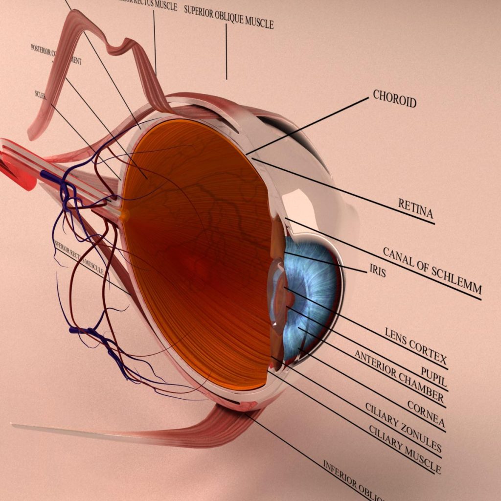
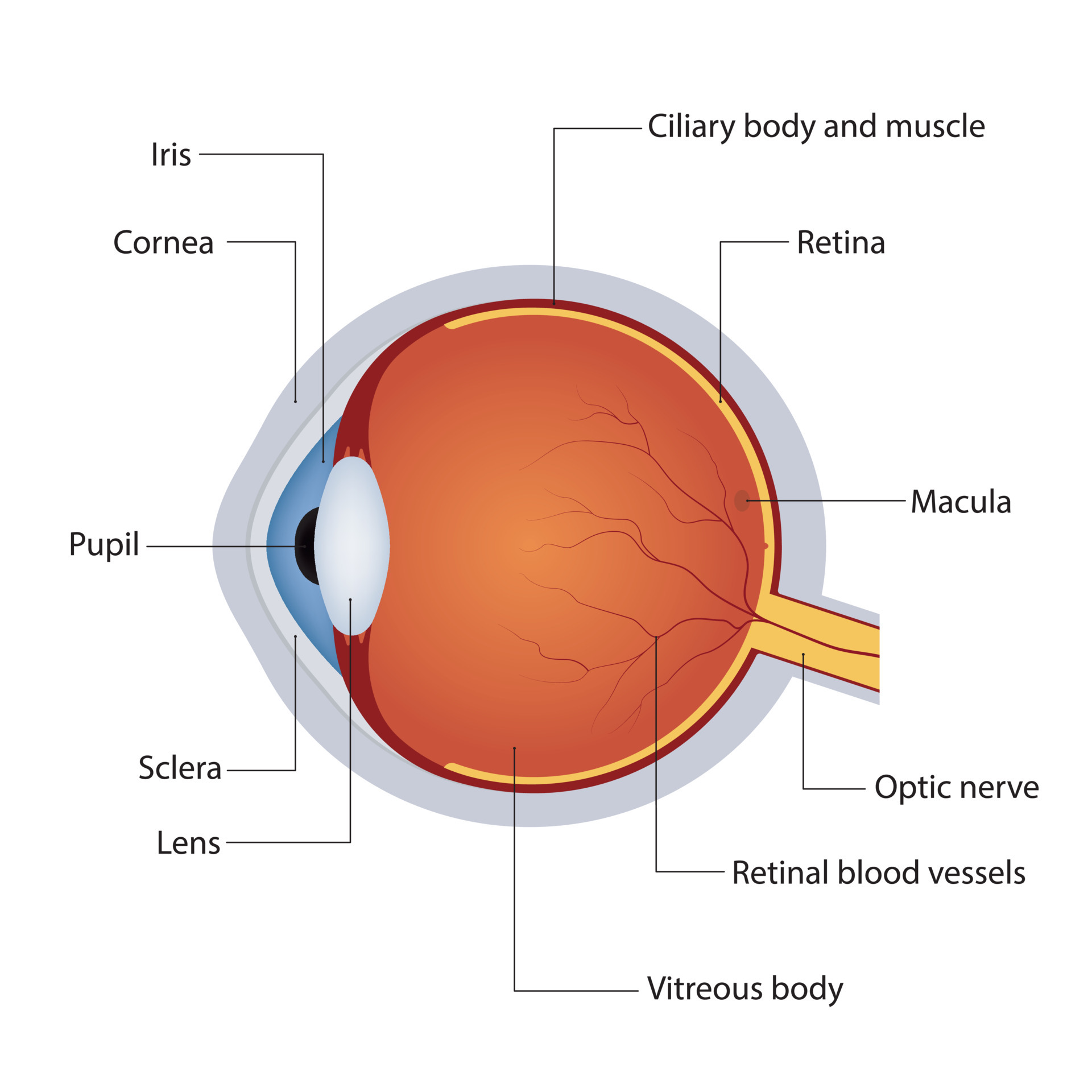
An anatomical model of the left eye with a cut section to display the layers of labelled anatomy. The eye sits in a protective bony socket called the orbit. A hole in the middle of the iris that changes size to let in more or less light. The eyeball, eye socket, brow ridge, eyelids, tear duct, sclera, iris, pupil, cornea, glabella, and epicanthic fold. The bottom part of the eyes has a slight lid. Use your pen to lightly make strokes in the outward direction under the eye. “cajal’s drawings of the retina are as beautiful as they are anatomically accurate. These interactive figures are provided for use in medical student education. Cells in the retina of the eye. The inner sensorineural layer is known as the retina.

Anatomy of the Human Eye
Start with a basic sketch. Web anatomy for artists: Pick the right drawing tools. Fascial sheath (tenon’s capsule) function. Get to know the eye structure.
Anatomy Human Eye Cross Section 3D Model Kezan's Portfolio
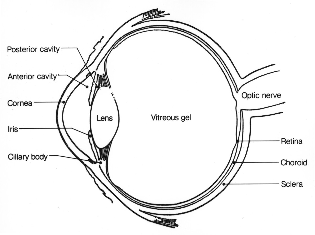
Study the diagram below or click here for an interactive study guide and game! For more video tutorials visit www.proko.com and subscribe to the newsletter. Use your pen to lightly make strokes in the outward direction under the eye. Start with a basic sketch. Why learn how to draw human anatomy.

Eye Anatomy
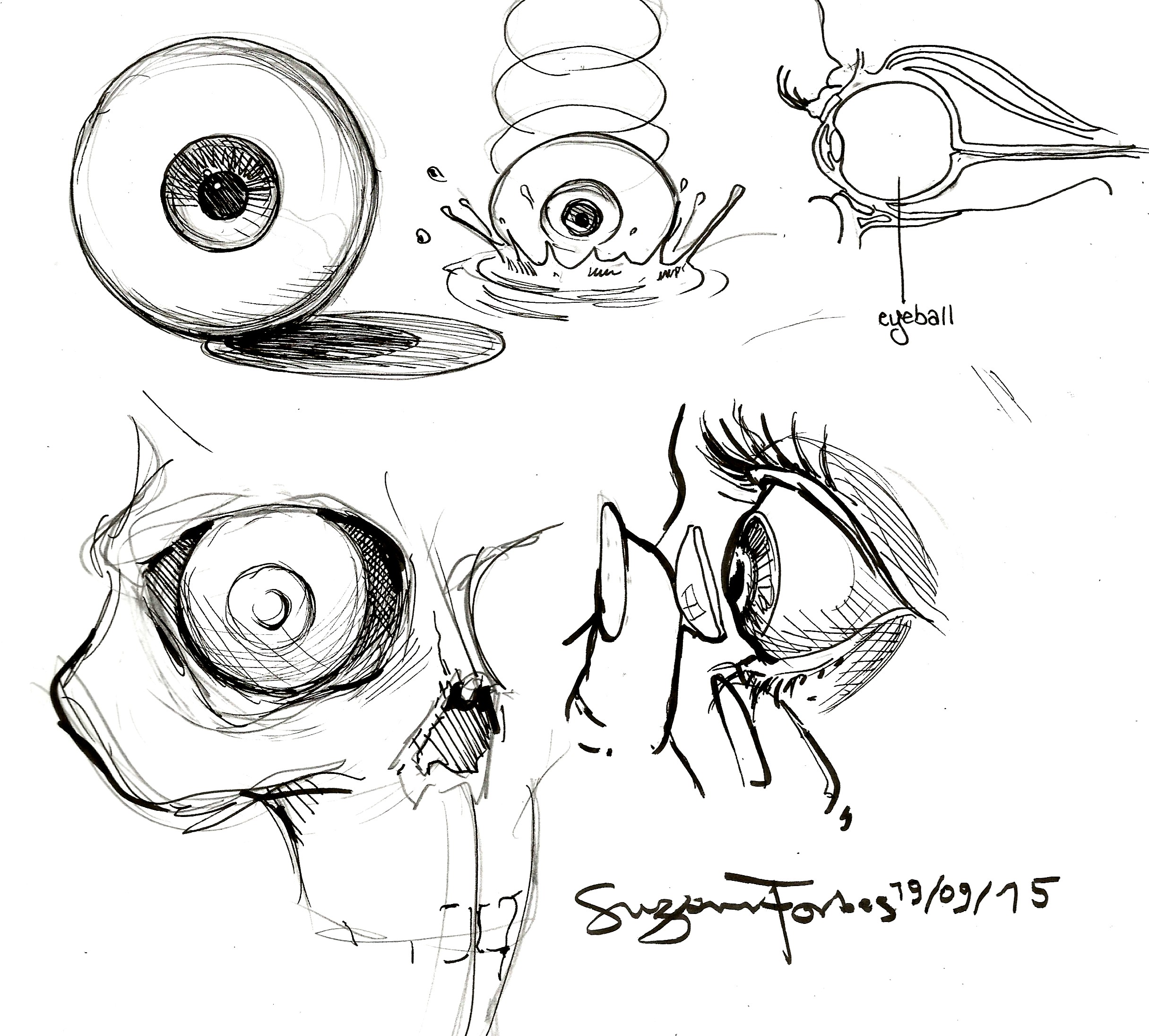
Broadly, the retina can be divided into the neural layer that has light receptors and deciphers the light stimuli and the pigmented area that absorbs light so that it does not reflect on the neural layer and create distortion. Web consider how the inner part of the eyes has lines that seem to push outward. Why learn how to draw.
/GettyImages-1128675065-e4bac15b0f39449dba31f25f1020bc8a.jpg)
Human Eye Diagram Eyeball Diagram Diagram Of The Eye Images and
Santiago ramón y cajal, 1904. Web in this video, we're going to talk about the structure of the eye. And i'm going to label is sclera. Retinal pigment epithelium (rpe) blood supply. Refractive media of the eyeball.
Drawing tutorial How to draw a perfect eye.
The bottom part of the eyes has a slight lid. Drawing digitally with layers makes finding the most compelling eyes for your character easier, as well. The eyeball, eye socket, brow ridge, eyelids, tear duct, sclera, iris, pupil, cornea, glabella, and epicanthic fold. Curved to bend light into your eye, its tough and clear like a windshield to protect your.
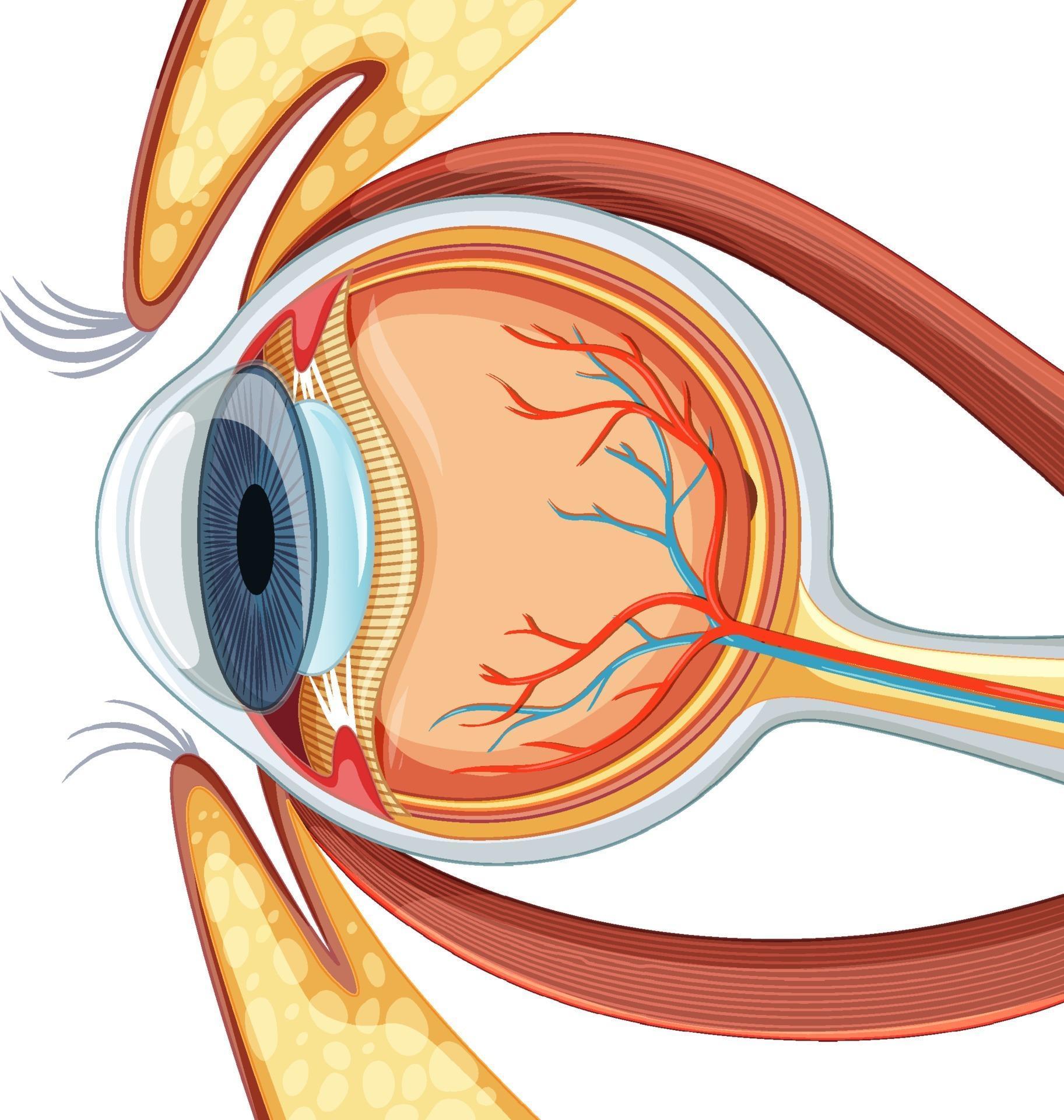
Diagram of human eyeball anatomy 3188538 Vector Art at Vecteezy
Santiago ramón y cajal, 1904. The bottom part of the eyes has a slight lid. “cajal’s drawings of the retina are as beautiful as they are anatomically accurate. Web anatomy for artists: Curved to bend light into your eye, its tough and clear like a windshield to protect your eye from dust.

Learn To Draw Eyes Drawing On Demand Anatomy sketches, Anatomy art
Web in this video, we're going to talk about the structure of the eye. Study the diagram below or click here for an interactive study guide and game! The eyeball, eye socket, brow ridge, eyelids, tear duct, sclera, iris, pupil, cornea, glabella, and epicanthic fold. And i'm going to label is sclera. Web in this helpful guide to eyes, i’ll.
Anatomy of the Eye Human Eye Anatomy Owlcation
Start with a basic sketch. Santiago ramón y cajal, 1904. Curved to bend light into your eye, its tough and clear like a windshield to protect your eye from dust. Here is a tour of the eye starting from the outside, going in through the front and working to the back. Retinal pigment epithelium (rpe) blood supply.
Structure of anatomy human eye. Detailed diagram of eyeball. Side view
Science & technology 3d models. Why learn how to draw human anatomy. Web diagram of the eye. Hi friends,in this video i will show you how to draw a eye diagram very easy way, its a step by step tutorial to draw diagram of eye anatomy. Web © 2023 google llc.
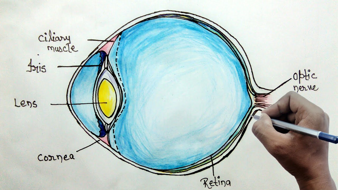
How to draw human eye diagram for beginners YouTube
The eyeball, eye socket, brow ridge, eyelids, tear duct, sclera, iris, pupil, cornea, glabella, and epicanthic fold. Anterior chamber angle and ciliary body. So i'm just drawing that in. Hi friends,in this video i will show you how to draw a eye diagram very easy way, its a step by step tutorial to draw diagram of eye anatomy. An anatomical.
Web Discovering The Underlying Anatomy Of The Eyeball Can Help You Bring Life And Feeling Into Your Drawings, From The Corners Of The Eye To The Pupils.
Fascial sheath (tenon’s capsule) function. And i'm going to label is sclera. “cajal’s drawings of the retina are as beautiful as they are anatomically accurate. Web diagram of the eye.
Retinal Pigment Epithelium (Rpe) Blood Supply.
Get to know the eye structure. The first thing we're going to draw is the white part of the eye, which is known as the sclera. External landmarks and extraocular muscles. The eyeball, eye socket, brow ridge, eyelids, tear duct, sclera, iris, pupil, cornea, glabella, and epicanthic fold.
An Anatomical Model Of The Left Eye With A Cut Section To Display The Layers Of Labelled Anatomy.
Web in this video, we're going to talk about the structure of the eye. Try to run your pen along that lid to demarcate where you should stop shading. Parts of the eye outside the eyeball. Cells in the retina of the eye.
Web Discovering The Underlying Anatomy Of The Eyeball Can Help You Bring Life And Feeling Into Your Drawings, From The Corners Of The Eye To The Pupils.
2.9m views 11 years ago #sketch #eyes #anatomy. Use your pen to lightly make strokes in the outward direction under the eye. The bottom part of the eyes has a slight lid. 318 views 1 year ago scientific diagram drawing tutorials.