Human Cell Drawing
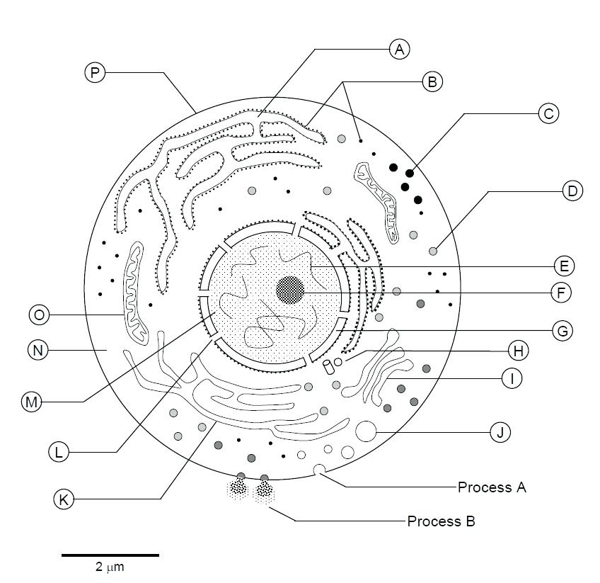
Human Cell Drawing - Diagram of the human cell illustrating the different parts of the cell. 4.9k views 11 months ago india. The top half of the cell volume was removed. This unit is part of the biology library. Other cells acquire specialized functions as they mature. Number 1 shows the nucleus, numbers 3 to 13 show different organelles immersed in the cytosol, and number 14 on the surface of the cell shows the plasma membrane. Most cells have only one nucleus, but some have more than one, and others—like mature red blood cells—don’t have one at all. The cell membrane is the outer coating of the cell and contains the cytoplasm, substances within it and the organelle. Web visual guide to human cells. What are the parts of a cell?
:) thanks for watching our channel. Number 1 shows the nucleus, numbers 3 to 13 show different organelles immersed in the cytosol, and number 14 on the surface of the cell shows the plasma membrane. What are the parts of a cell? Other cells acquire specialized functions as they mature. The top half of the cell volume was removed. In this drawing,i will show you how to draw and label a simple human cell easy step by step for beginners. The cell membrane is the outer coating of the cell and contains the cytoplasm, substances within it and the organelle. Web accessible resource database of images, videos, and animations of cells, capturing a wide diversity of organisms, cell types, and cellular processes. Web diagram of the human cell illustrating the different parts of the cell. Let’s learn about cell parts.
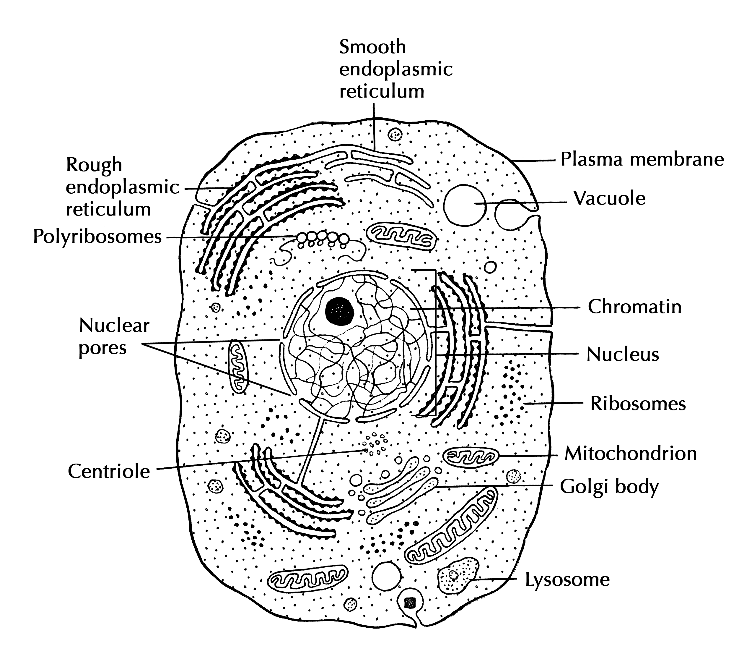
The cell membrane is the outer coating of the cell and contains the cytoplasm, substances within it and the organelle. To learn more about cells and cell parts, visit building blocks of life for more of the story. Let’s learn about cell parts. The atlas is likely to lead to major advances in the way illnesses are diagnosed and treated. In this drawing,i will show you how to draw and label a simple human cell easy step by step for beginners. Human cell diagram, human cell drawing labeled,. :) thanks for watching our channel. What is your request drawing? The interior of human cells is divided into the nucleus and the cytoplasm. Free for commercial use high quality images.

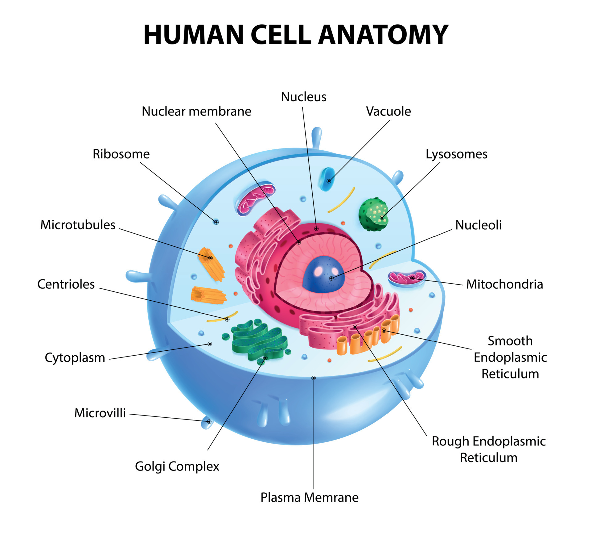
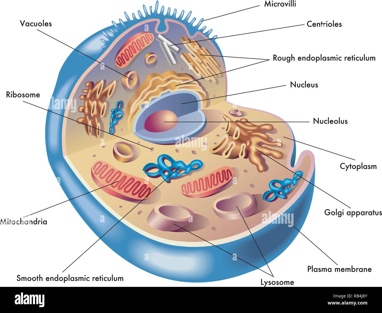
Human cell anatomy and organels diagram
Web visual guide to human cells. Human cell diagram, human cell drawing labeled,. What are the parts of a cell? This unit is part of the biology library. To learn more about cells and cell parts, visit building blocks of life for more of the story.

69,023 Human Cell Structure Images, Stock Photos & Vectors Shutterstock
Web learn the similarities and differences in the anatomy of animal, plant, fungal, and bacterial cell types by exploring our cell viewer. Free for commercial use high quality images. The top half of the cell volume was removed. Within the nucleus is a spherical body known as the nucleolus, which contains clusters of protein, dna, and rna. :) thanks for.

How to Draw Human Cell Step by Step YouTube
Number 1 shows the nucleus, numbers 3 to 13 show different organelles immersed in the cytosol, and number 14 on the surface of the cell shows the plasma membrane. :) thanks for watching our channel. Dna sequencing data processing genetic genomic analysis. Other cells acquire specialized functions as they mature. Web interactive guide to stem cells and cell biology with.
Human Cell Drawing at Explore collection of Human
Test your knowledge of the parts of a cell with our unlabeled worksheet. Learn faster with interactive cell quizzes. Most cells have only one nucleus, but some have more than one, and others—like mature red blood cells—don’t have one at all. What are the parts of a cell? The cell membrane is the outer coating of the cell and contains.
Human Cells
Skin tissue cancerous cells, melanoma. Web page 1 of 100. :) thanks for watching our channel. Test your knowledge of the parts of a cell with our unlabeled worksheet. The atlas is likely to lead to major advances in the way illnesses are diagnosed and treated.
Human Cell Sketch at Explore collection of Human
Other cells acquire specialized functions as they mature. Web although the map covers just a fraction of the organ—a whole brain is a million times larger—that piece contains roughly 57,000 cells, about 230 millimeters of blood vessels, and nearly 150. Web visual guide to human cells. Web diagram of the human cell illustrating the different parts of the cell. Browse.
Medical illustration of elements of human cell Stock Vector Image & Art
Cells contain parts called organelles. Web interactive guide to stem cells and cell biology with 3d models and real microscopy data of gfp labeled hipscs. 12k views 6 months ago drawing for. The atlas is likely to lead to major advances in the way illnesses are diagnosed and treated. 4.9k views 11 months ago india.

Human Cell Diagram, Parts, Pictures, Structure and Functions
Skin tissue cells, layers of skin, blood in vein. What are the parts of a cell? Web diagram of the human cell illustrating the different parts of the cell. Web the nucleus is a large organelle that contains the cell’s genetic information. Cells contain parts called organelles.

The cell structure. Organelles. Watercolor. Kateryna Tonyuk Cell
12k views 6 months ago drawing for. The top half of the cell volume was removed. Web page 1 of 100. Web diagram of the human cell illustrating the different parts of the cell. Cross section animal cell structure detailed colorful anatomy.

3d human cell
Web a cubic millimeter of brain tissue may not sound like much. This unit is part of the biology library. 99,000+ vectors, stock photos & psd files. Browse videos, articles, and exercises by topic. Skin tissue cancerous cells, melanoma.
All Cells Have A Cell Membrane That Separates The Inside And The Outside Of The Cell, And Controls What Goes In And Comes Out.
What are the parts of a cell? The cell membrane is the outer coating of the cell and contains the cytoplasm, substances within it and the organelle. Eukaryotic cell diagram, vector illustration, text on own layer. The interior of human cells is divided into the nucleus and the cytoplasm.
Web The Nucleus Is A Large Organelle That Contains The Cell’s Genetic Information.
Web figure 4.1.1 4.1. 4.9k views 11 months ago india. Other cells acquire specialized functions as they mature. Web how to draw human cell step by step.
Web Browse 5,900+ Human Cell Diagram Stock Photos And Images Available, Or Search For Cells To Find More Great Stock Photos And Pictures.
Learn faster with interactive cell quizzes. The top half of the cell volume was removed. Free for commercial use high quality images. This unit is part of the biology library.
Number 1 Shows The Nucleus, Numbers 3 To 13 Show Different Organelles Immersed In The Cytosol, And Number 14 On The Surface Of The Cell Shows The Plasma Membrane.
99,000+ vectors, stock photos & psd files. 12k views 6 months ago drawing for. Most cells have only one nucleus, but some have more than one, and others—like mature red blood cells—don’t have one at all. Web interactive guide to stem cells and cell biology with 3d models and real microscopy data of gfp labeled hipscs.