Human Heart Drawing Labeled
Human Heart Drawing Labeled - This tool provides access to several medical illustrations, allowing the user to interactively discover heart anatomy. Blood brings oxygen and nutrients to your cells. The two upper chambers are called the atria, the remaining two lower chambers are the ventricles. It rests on the diaphragm, the muscular partition between the chest and the abdominal cavity. Web 1.3m views 3 years ago 3 products. In this interactive, you can label parts of the human heart. Includes an exercise, review worksheet, quiz, and model drawing of an anterior vi [right atrium and ventricle of the heart (labeled)] Your heart is in the center of your chest, near your lungs. Rotate the 3d model to see how the heart's valves control blood flow between heart chambers and blood flow out of the heart.
Take a look at our labeled heart diagrams (see below) to get an overview of all of the parts of the heart. Find a piece of paper and something to draw with. The two upper chambers are called the atria, the remaining two lower chambers are the ventricles. The heart is a mostly hollow, muscular organ composed of cardiac muscles and connective tissue that acts. Pencil sketch of the human heart. The two upper chambers are called the left and the right atria, and the two lower chambers are known as the left and the right ventricles. The human heart is located within the thoracic cavity, medially between the lungs in the space known as the mediastinum. Right atrium, left atrium, right ventricle and left ventricle. Drag and drop the text labels onto the boxes next to the diagram. Anatomy and function of the heart.
Web heart pictures, diagram & anatomy | body maps. Relate the structure of the heart to its function as a pump. Web inside, the heart is divided into four heart chambers: 244 × 240 pixels | 489 × 480 pixels | 782 × 768 pixels | 1,043 × 1,024 pixels | 2,086 × 2,048 pixels | 663 × 651 pixels. Web the human heart is primarily comprised of four chambers. Web 1.3m views 3 years ago 3 products. Embark on a journey into the beauty of human anatomy with a mesmerizing pencil sketch of the human heart. The two upper chambers are called the atria, the remaining two lower chambers are the ventricles. Web + show all. At the heart of it all:
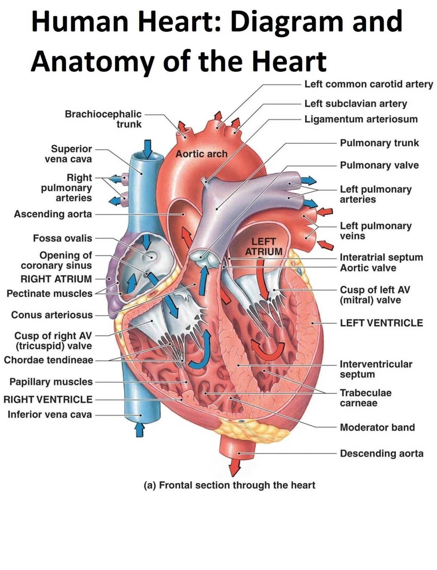
The Human Heart Diagram Display Poster Diagram and Anatomy of the Heart
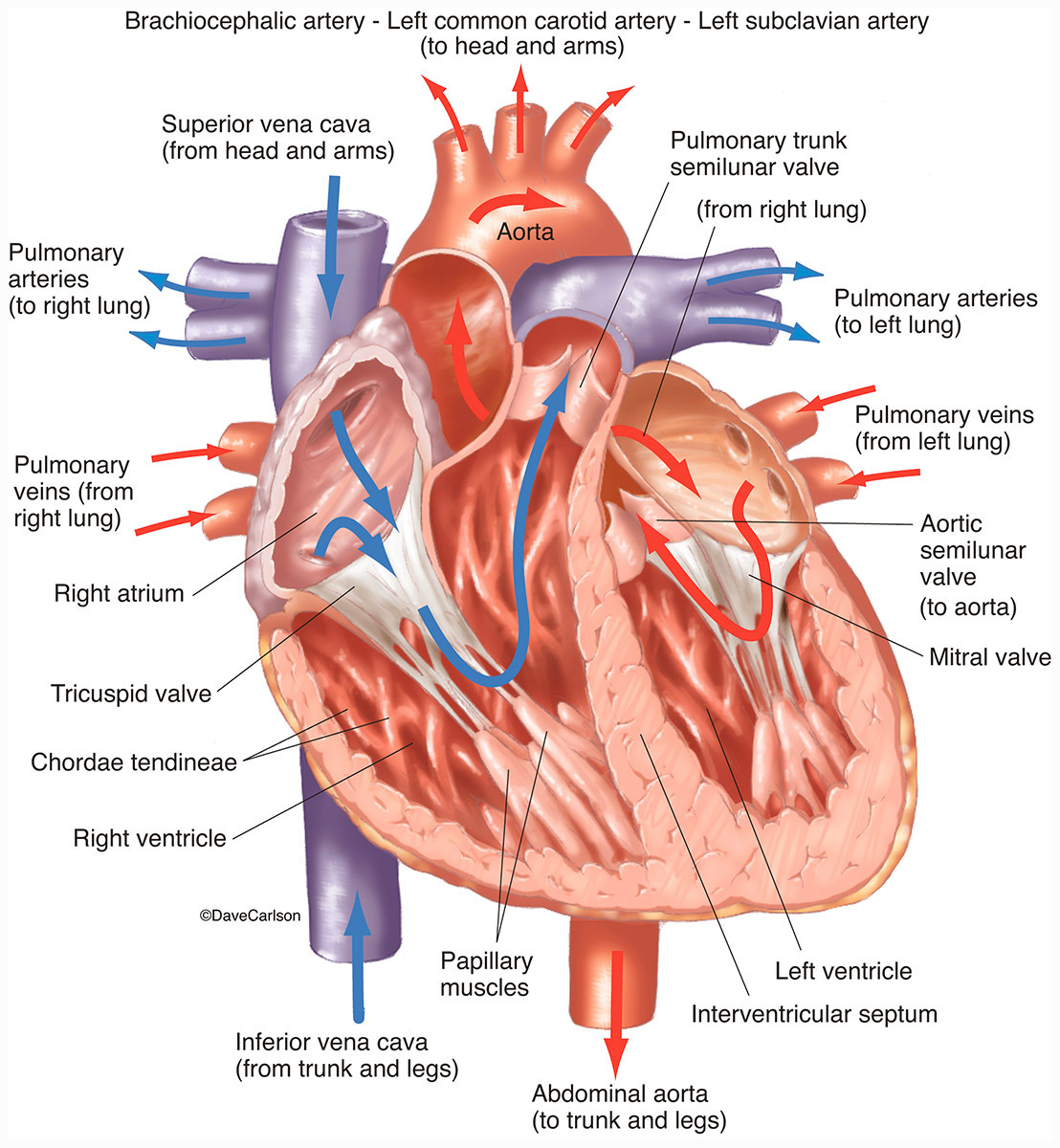
Web heart pictures, diagram & anatomy | body maps. Start with the pulmonary veins. To find a good diagram, go to google images, and type in the internal structure of the human heart. Web in order to understand how that happens, it is necessary to understand the anatomy and physiology of the heart. Controls the rhythm and speed of your.

How to Draw the Internal Structure of the Heart 14 Steps
Start with the pulmonary veins. 93 kb) render this image in. Neatly print the names around your drawing and then use a ruler to draw an arrow to the corresponding part. Rotate the 3d model to see how the heart's valves control blood flow between heart chambers and blood flow out of the heart. Blood brings oxygen and nutrients to.
.svg/1043px-Diagram_of_the_human_heart_(cropped).svg.png)
FileDiagram of the human heart (cropped).svg Wikipedia
The chambers are separated by heart valves, which make sure that the blood keeps flowing in the right direction. Your heart is in the center of your chest, near your lungs. Web the human heart is primarily comprised of four chambers. Anatomy and function of the heart. Find a piece of paper and something to draw with.

Anatomy and Physiology Heart Anatomy
Take a look at our labeled heart diagrams (see below) to get an overview of all of the parts of the heart. Web the heart | circulatory anatomy. Read more about heart valves and how they help blood flow through the heart. Two atria (right and left) and two ventricles (right and left). Find an image that displays the entire.

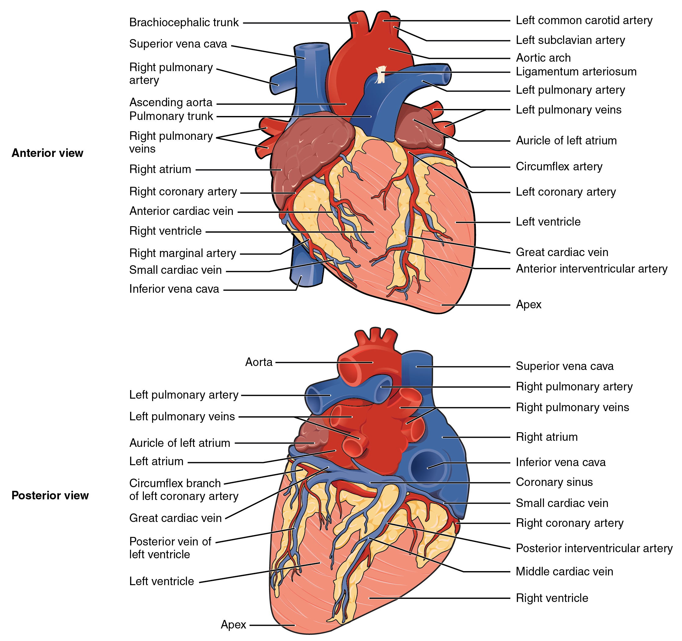
Heart Anatomy chambers, valves and vessels Anatomy & Physiology
Once you’re feeling confident, you can test yourself using the unlabeled diagrams of the parts of the heart below. Web in this lecture, dr mike shows the two best ways to draw and label the heart! The bottom tip of the heart, known as its apex, is turned to the left, so that about 2/3 of the heart is located.

Human heart anatomy. Vector diagram in 2021 Heart anatomy, Human
This artwork delves into the depths of medical illustration, celebrating the complexity and elegance of the heart's form. Size of this png preview of this svg file: It consists of four chambers, four valves, two main arteries (the coronary arteries), and the conduction system. Start with the pulmonary veins. Web anatomy of the human heart.
Human Heart Front View Interior Carlson Stock Art
Read more about heart valves and how they help blood flow through the heart. 93 kb) render this image in. It also takes away carbon dioxide and other waste so other organs can dispose of them. Find an image that displays the entire heart, and click on it to enlarge it. Controls the rhythm and speed of your heart rate.

humanheartdiagram Tim's Printables
Size of this png preview of this svg file: Original file (svg file, nominally 663 × 651 pixels, file size: 93 kb) render this image in. Web label the parts of the heart to reference it for anatomy. This tool provides access to several medical illustrations, allowing the user to interactively discover heart anatomy.
19.1 Heart Anatomy Anatomy and Physiology
Find an image that displays the entire heart, and click on it to enlarge it. Web 1.3m views 3 years ago 3 products. The two upper chambers are called the atria, the remaining two lower chambers are the ventricles. Web your heart’s main function is to move blood throughout your body. This artwork delves into the depths of medical illustration,.
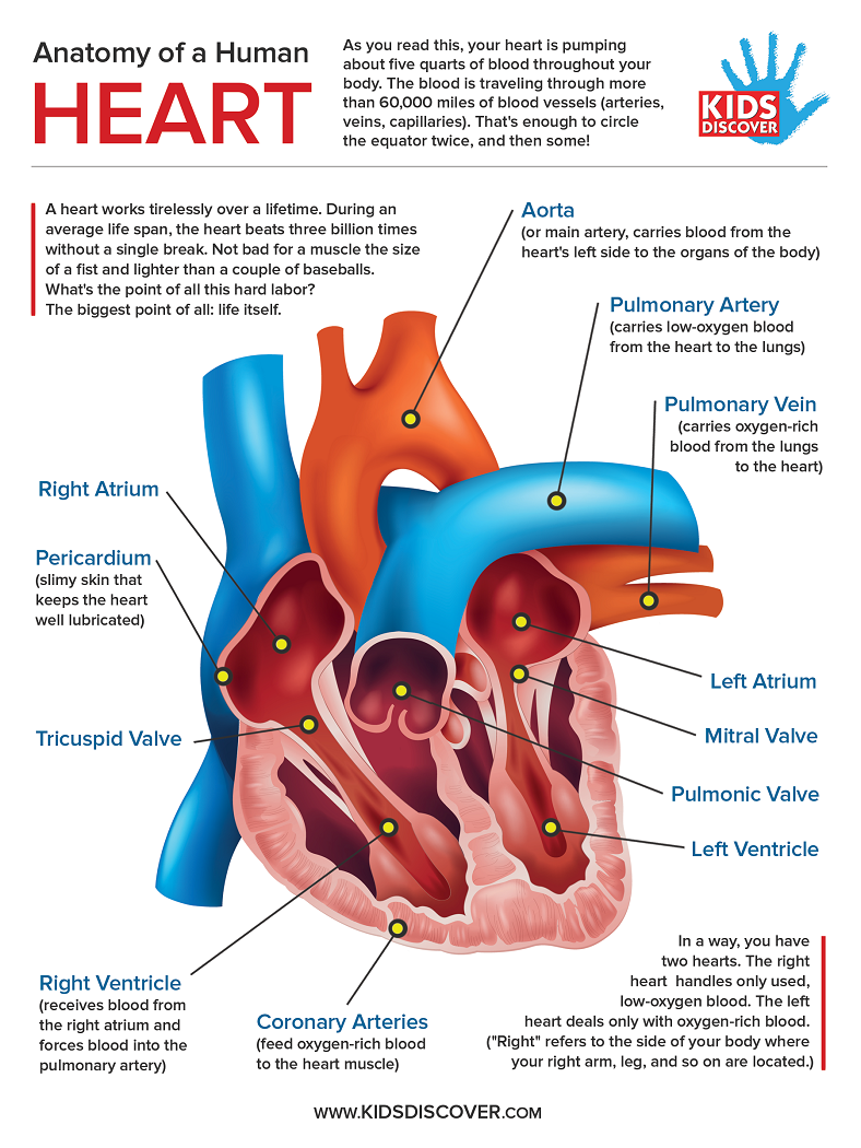
Heart Labeled Anatomy
It rests on the diaphragm, the muscular partition between the chest and the abdominal cavity. Web your heart’s main function is to move blood throughout your body. Web in this lecture, dr mike shows the two best ways to draw and label the heart! Figure 19.2 shows the position of the heart within the thoracic cavity. Two atria (right and.
Anatomy And Function Of The Heart.
It has four hollow chambers surrounded by muscle and other heart tissue. Web the heart is located in the thoracic cavity medial to the lungs and posterior to the sternum. Read more about heart valves and how they help blood flow through the heart. Web your heart’s main function is to move blood throughout your body.
Size Of This Png Preview Of This Svg File:
Web cardiovascular system anatomy. Identify the tissue layers of the heart. Right atrium, left atrium, right ventricle and left ventricle. It rests on the diaphragm, the muscular partition between the chest and the abdominal cavity.
The Heart Is A Muscular Pumping Organ Located Medial To The Lungs Along The Body’s Midline In The Thoracic Region.
Original file (svg file, nominally 663 × 651 pixels, file size: Embark on a journey into the beauty of human anatomy with a mesmerizing pencil sketch of the human heart. Compare systemic circulation to pulmonary circulation. In humans, the heart is situated between the two lungs and slightly to the left of center, behind the breastbone.
Web Anatomy Of The Human Heart And Coronaries:
Web describe the internal and external anatomy of the heart. Web + show all. This artwork delves into the depths of medical illustration, celebrating the complexity and elegance of the heart's form. It consists of four chambers, four valves, two main arteries (the coronary arteries), and the conduction system.