Integumentary System Drawing With Label
Integumentary System Drawing With Label - Beneath the dermis lies the hypodermis, which is composed mainly of loose. Web the integumentary system is the body's outermost layer. A drawing of a person with an area of exposed skin (like an arm) with the label integument 2. Bone tissue and the skeletal system. The drawing and anatomical labeling of these illustrations was done by b. Web select the correct structure found in the integumentary system. In humans, this system accounts. The dermis, the epidermis, the erector pili muscle, hair follicles, the hypodermis, meissner's. Absorbs and helps heal abrasions, cuts and other injuries. Exercise 2 layers of epidermis.
Exercise 2 layers of epidermis. It comprises the skin and its appendages, which act as a physical barrier between the external environment and the internal environment that it serves to protect and maintain the body of the animal. Web select the correct structure found in the integumentary system. Most skin disorders are relatively benign, but a few, including melanomas, can be fatal if untreated. It works to protect the body from harm and maintain homeostasis by working with other bodily systems. 5.3 functions of the integumentary system. Most of the skin can be classified as thin skin. Integumentary system labeling — quiz information. It also helps retain bodily fluids, eliminate waste products, and regulate body temperature. Sketch the skin and label the parts of the integument shown in figure 5.2 above, observed at low and high magnification.
Image a on each chart is for reference! It is the system that can instantly tell us whether someone is young or old, someone’s. 5.3 functions of the integumentary system. 15 questions on the skin : The skin protects deeper tissues from mechanical damage (bumps), chemical damage (acids and. Web official ninja nerd website: Sketch the skin and label the parts of the integument shown in figure 5.2 above, observed at low and high magnification. Web the integumentary system is the body's outermost layer. One of the best ways to start learning about a new system, organ or region is with a labeled diagram showing you all of the main structures found within it. Redraw and label image b below.

Diagrams The integumentary system
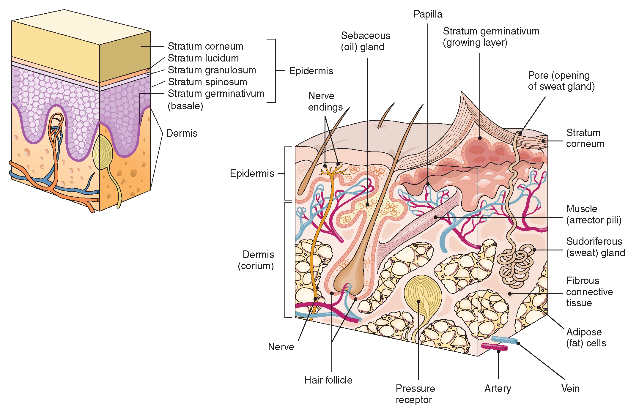
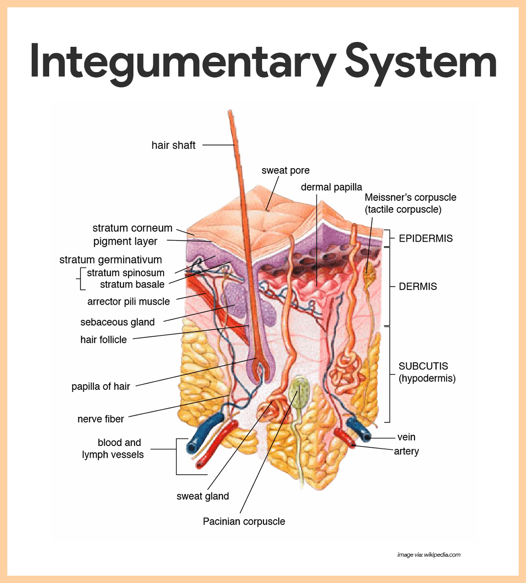
Web the integumentary system, or skin, is the largest organ in the body. From deep to superficial, these layers are the stratum basale, stratum spinosum, stratum granulosum, and stratum corneum. 15 questions on the skin : It is the system that can instantly tell us whether someone is young or old, someone’s. 5.2 accessory structures of the skin.
Integumentary system diagram
If you look in the mirror you see it, if you look anywhere on your body you see and if you look around you in the outside world, you see it. In humans, this system accounts. Mainly it is the body's outer skin. The dermis, the epidermis, the erector pili muscle, hair follicles, the hypodermis, meissner's. 5.1 layers of the.
The Integumentary System (Structure and Function) (Nursing) Part 1
Web the integumentary system is the set of organs forming the outermost layer of an animal's body. 5.1 layers of the skin. Web the integumentary system, or skin, is the largest organ in the body. Mainly it is the body's outer skin. This article digs into the specifics about.

The Skin
Web tutorial on drawing anatomical structures. Integumentary system histology the cell and tissue structures of the integumentary system are suited for the functions performed. Mainly it is the body's outer skin. If you look in the mirror you see it, if you look anywhere on your body you see and if you look around you in the outside world, you.
Integumentary System Anatomy and Physiology Nurseslabs
The first thing a clinician sees is the skin, and so the examination of the skin should be part of any thorough physical examination. This drawing and labeling activity will help you internalize those layers. A couple of the more noticeable disorders, albinism and vitiligo, affect the appearance. Integumentary system histology the cell and tissue structures of the integumentary system.
The Human Skin The largest organ of the Integumentary System HubPages
The first thing a clinician sees is the skin, and so the examination of the skin should be part of any thorough physical examination. Redraw and label image b below. The functions of the integumentary system are: Web skin that has four layers of cells is referred to as “thin skin.”. For nursing students eager to grasp the anatomy and.

Integumentary System Parts And Their Functions
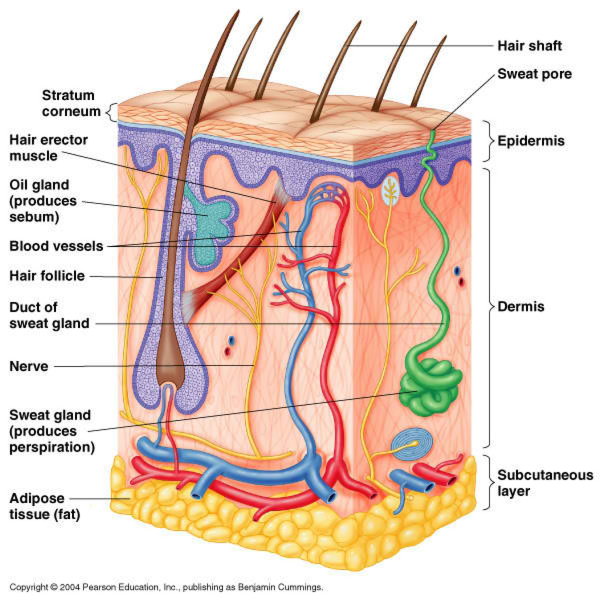
Exercise 2 layers of epidermis. Besides the skin, it comprises the hair and nails as well, which are appendages of the skin. For nursing students eager to grasp the anatomy and physiology of our first line of defense. Web your integumentary system stores fat, water, glucose and vitamin d, and helps support your immune system to protect you from diseases..
Integumentary system parts Quizzes and diagrams Kenhub
Web the integumentary system, or skin, is the largest organ in the body. Web the integumentary system is the body system which surrounds you, both literally and metaphorically speaking. Beneath the dermis lies the hypodermis, which is composed mainly of loose. Anatomy atlas of the integument (skin, hair, skin glands). Web skin that has four layers of cells is referred.
:max_bytes(150000):strip_icc()/skin_structure-592308b15f9b58f4c0153a00.jpg)
The Layers of the Integumentary System
Web unlock the mysteries of our skin and its allies with the integumentary system guide; Exercise 2 layers of epidermis. Skin w/o hair using colored pens/pencils, draw the histology image b from the “skin w/o hair” chart in the space below. The first thing a clinician sees is the skin, and so the examination of the skin should be part.
Integumentary System Anatomy & Physiology
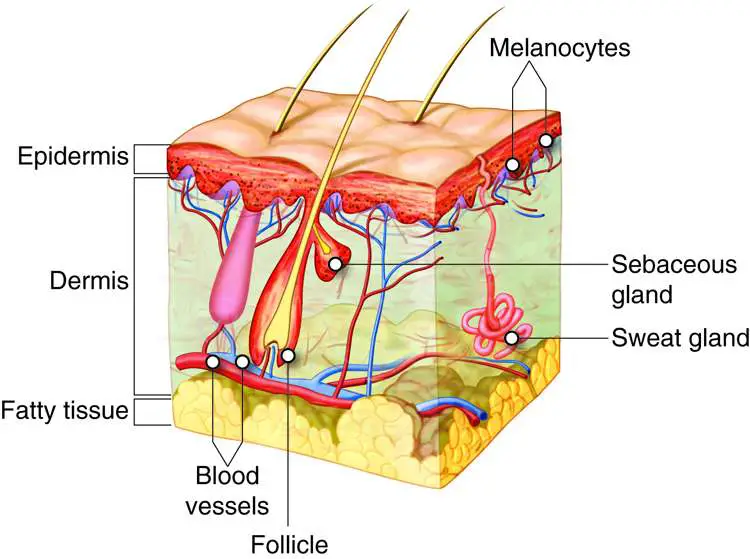
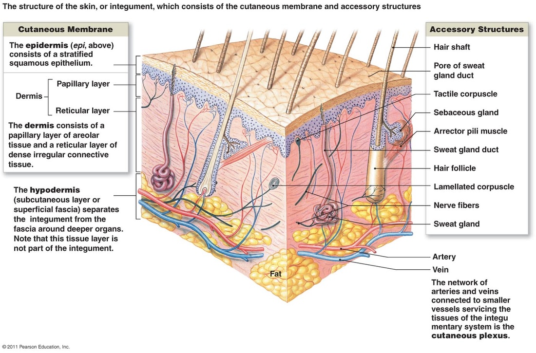
The skin and its accessory structures make up the integumentary system, which provides the body with overall protection. The epidermis, made of closely packed epithelial cells, and the dermis, made of dense, irregular connective tissue that houses blood vessels, hair follicles, sweat glands, and other structures. For nursing students eager to grasp the anatomy and physiology of our first line.
Besides The Skin, It Comprises The Hair And Nails As Well, Which Are Appendages Of The Skin.
The dermis, the epidermis, the erector pili muscle, hair follicles, the hypodermis, meissner's. Web the integumentary system is the set of organs forming the outermost layer of an animal's body. Most of the skin can be classified as thin skin. One of the best ways to start learning about a new system, organ or region is with a labeled diagram showing you all of the main structures found within it.
Redraw And Label Image B Below.
The skin is composed of two main layers: 15 questions on the skin : Web unlock the mysteries of our skin and its allies with the integumentary system guide; Integumentary system labeling — quiz information.
The Functions Of The Integumentary System Are:
The drawing and anatomical labeling of these illustrations was done by b. From deep to superficial, these layers are the stratum basale, stratum spinosum, stratum granulosum, and stratum corneum. Your integumentary system has many important functions. “thick skin” is found only on the palms of the hands and the soles of the feet.
Sketch The Skin And Label The Parts Of The Integument Shown In Figure 5.2 Above, Observed At Low And High Magnification.
It comprises the skin and its appendages, which act as a physical barrier between the external environment and the internal environment that it serves to protect and maintain the body of the animal. This article digs into the specifics about. Web study with quizlet and memorize flashcards containing terms like epidermis, dermis, subcutaneous layer and more. Skin w/o hair using colored pens/pencils, draw the histology image b from the “skin w/o hair” chart in the space below.