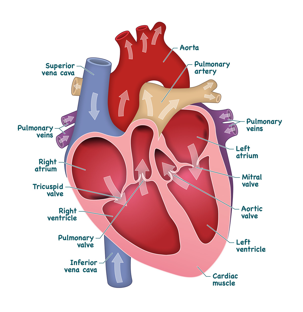
Labelled Heart Drawing
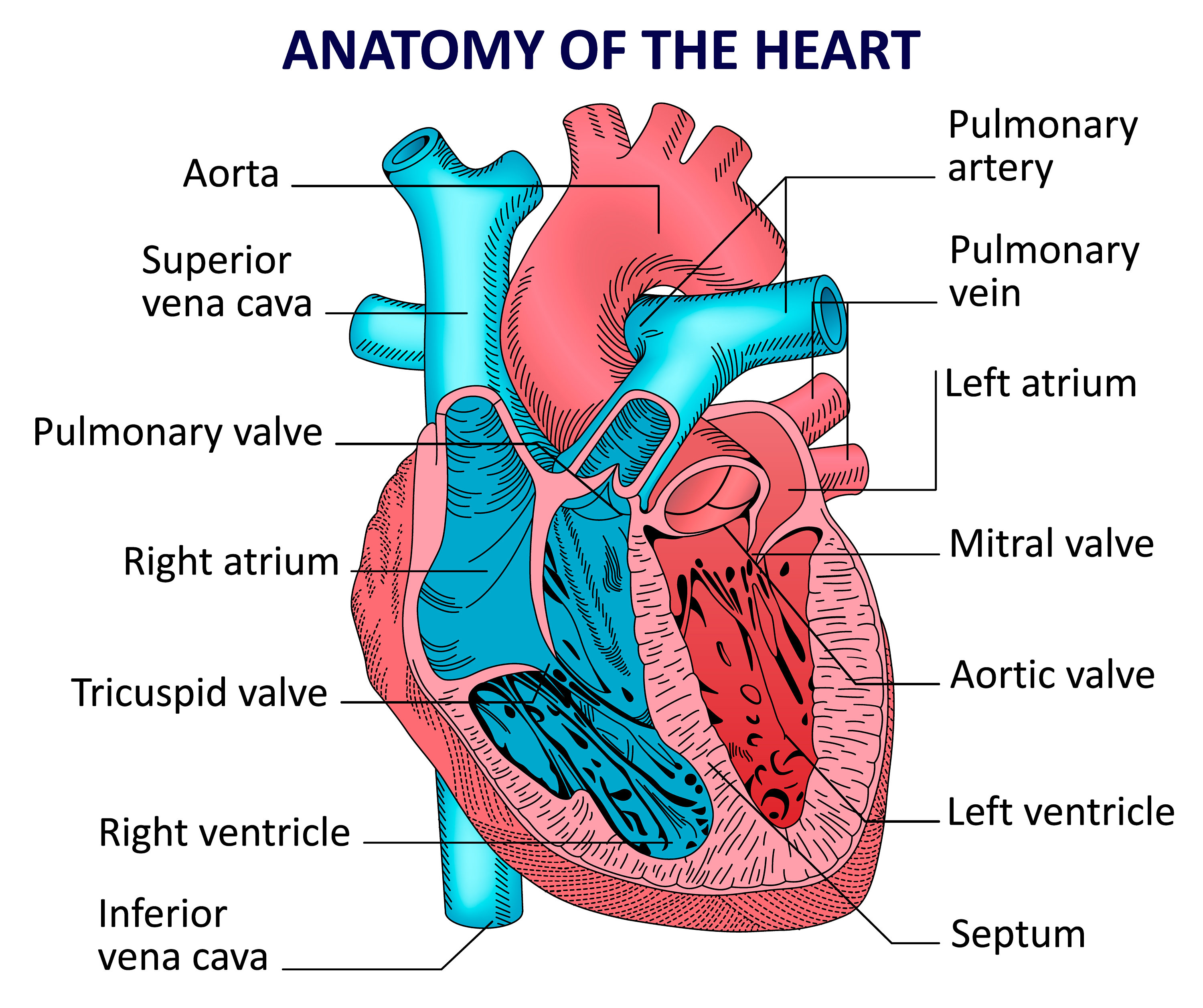
Labelled Heart Drawing - The right and left sides of the heart are separated by a muscle called the “septum.”. Web anatomy of the heart made easy along with the blood flow through the cardiac structures, valves, atria, and ventricles. Blood transports oxygen and nutrients to the body. The heart is a muscular organ that pumps blood through the blood vessels of the circulatory system. Size of this png preview of this svg file: Base (posterior), diaphragmatic (inferior), sternocostal (anterior), and left and right pulmonary surfaces. Web the heart is located in the thoracic cavity medial to the lungs and posterior to the sternum. Web step 1 and 6 involve a blood vessel, which makes sense as this is how blood enters and exits that side of the heart. Web anatomy of the heart: On its superior end, the base of the heart is attached to the aorta,mycontentbreak pulmonary arteries and veins, and the vena cava.
Size of this png preview of this svg file: Web anatomy of the heart made easy along with the blood flow through the cardiac structures, valves, atria, and ventricles. Base (posterior), diaphragmatic (inferior), sternocostal (anterior), and left and right pulmonary surfaces. Myocardium is the thick middle layer of muscle that allows your heart chambers to contract and relax to pump blood to your body.; The two upper chambers are called the atria, the remaining two lower chambers are the ventricles. The document said archewell had given out $3 million in grants. This strong muscle tissue powers the heart’s pumping action. The inferior tip of the heart, known as the apex, rests just superior to the diaphragm. Web by the end of this section, you will be able to: The heart is a muscular organ that pumps blood through the blood vessels of the circulatory system.
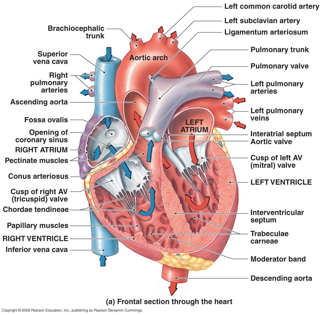
Your heart contains four muscular sections ( chambers) that briefly hold blood before moving it. Web discussed in this video is how to draw and label the structures of the heart, the layers of the heart, and a discussion on how blood flows within the heart. Blood transports oxygen and nutrients to the body. Web the most common heart attack symptoms or warning signs are chest pain, breathlessness, nausea, sweating etc. Create a “w” on top of the “j” shape to form the narrow tubes. Both sides work together to efficiently circulate the blood. On its superior end, the base of the heart is attached to the aorta,mycontentbreak pulmonary arteries and veins, and the vena cava. Web the heart is made of three layers of tissue. Web diagram of the human heart (cropped).svg. Web the heart has three layers.
Heart And Labels Drawing at GetDrawings Free download
Web labeled heart diagram showing the heart from anterior unlabeled heart diagrams (free download!) worksheet showing unlabelled heart diagrams. This thick layer is the muscle that contracts to pump and propel blood. Web step 1 and 6 involve a blood vessel, which makes sense as this is how blood enters and exits that side of the heart. It is also.

How to Draw the Internal Structure of the Heart 14 Steps
Both sides work together to efficiently circulate the blood. Web the bulk of its income came from one individual donor. 93 kb) render this image in. Endocardium is the thin inner lining of the heart chambers and also forms the surface of the valves.; Web labeled heart diagram showing the heart from anterior unlabeled heart diagrams (free download!) worksheet showing.

11+ Heart Drawing With Labels Robhosking Diagram
Cardiovascular system animation for u. It may be a straight tube, as in spiders and annelid worms, or a somewhat more elaborate structure with one or more receiving chambers (atria) and a main pumping chamber (ventricle), as in mollusks. Describe the internal and external anatomy of the heart. Drag and drop the text labels onto the boxes next to the.
Labeled Drawing Of The Heart at GetDrawings Free download
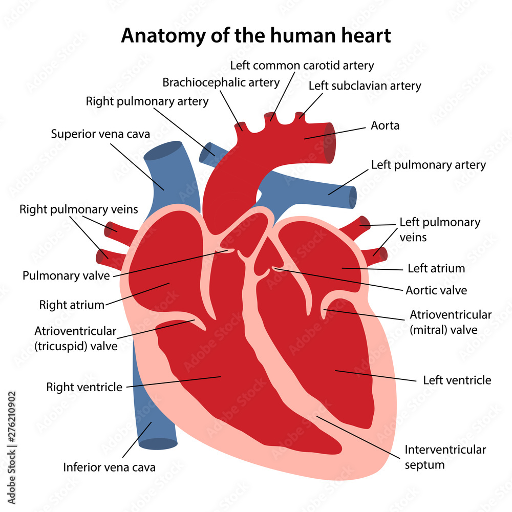
Web the human heart is primarily comprised of four chambers. Web in this lecture, dr mike shows the two best ways to draw and label the heart! In this interactive, you can label parts of the human heart. Web the heart is made of three layers of tissue. Web the cardiovascular system consists of the heart, blood vessels, and the.
When one teaches, two learn. The heart and the circulatory system
Both sides work together to efficiently circulate the blood. Pericardium is the sac that surrounds your heart. Your brain and nervous system direct your heart’s function. The inferior tip of the heart, known as the apex, rests just superior to the diaphragm. Web muscle and tissue make up this powerhouse organ.
Anatomy of the human heart. Cross sectional diagram of the heart with
Start your sketch at the edge of the superior vena cava, and work the shape down to the top edge of the heart’s body. Web the heart is made of three layers of tissue. Endocardium is the thin inner lining of the heart chambers and also forms the surface of the valves.; Web the epicardium covers the heart, wraps around.

How to Draw the Internal Structure of the Heart 13 Steps
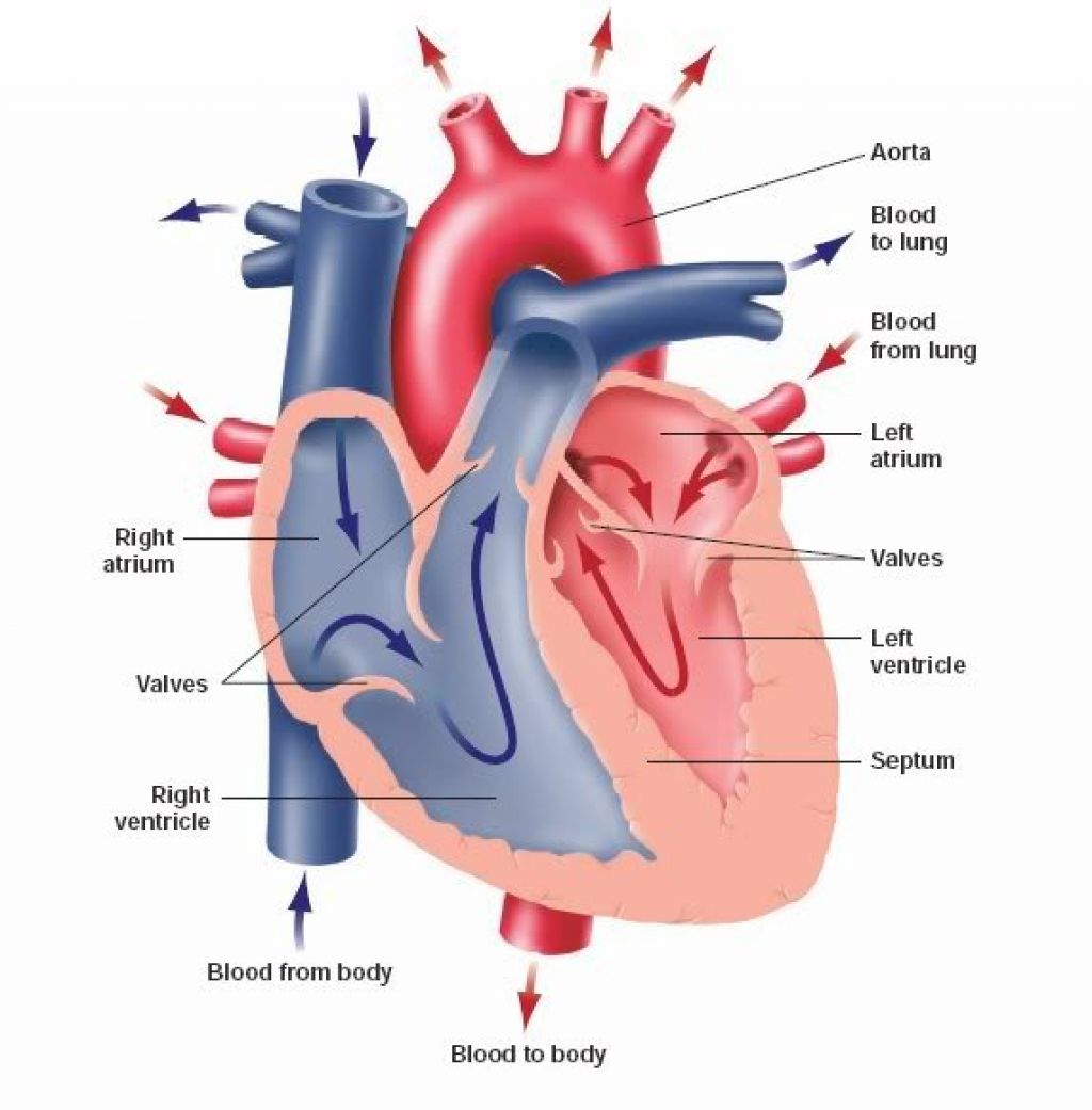
So if you remember this general pattern, it will help you recall the order in which blood flows through each side of the heart. In fishes the heart is a folded tube, with three or four enlarged areas that. Both sides work together to efficiently circulate the blood. The inferior tip of the heart, known as the apex, rests just.
The Human Heart Diagram Labeled
In fishes the heart is a folded tube, with three or four enlarged areas that. The right and left sides of the heart are separated by a muscle called the “septum.”. It is also involved in the removal of. 93 kb) render this image in. Base (posterior), diaphragmatic (inferior), sternocostal (anterior), and left and right pulmonary surfaces.

Cardiac cycle and the Human Heart A* understanding for iGCSE Biology 2
The heart is a muscular organ that pumps blood through the blood vessels of the circulatory system. Both sides work together to efficiently circulate the blood. The user can show or hide the anatomical labels which provide a useful tool to create illustrations perfectly adapted for teaching. The right and left sides of the heart are separated by a muscle.

heart anatomy labeling
Electrical impulses make your heart beat, moving blood through these chambers. 93 kb) render this image in. Describe the location and position of the heart within the body cavity. The inferior tip of the heart, known as the apex, rests just superior to the diaphragm. The two upper chambers are called the atria, the remaining two lower chambers are the.
The Two Upper Chambers Are Called The Atria, The Remaining Two Lower Chambers Are The Ventricles.
Web diagram of the human heart (cropped).svg. This shape represents the aorta. Myocardium is the thick middle layer of muscle that allows your heart chambers to contract and relax to pump blood to your body.; Relate the structure of the heart to its function as a pump.
Endocardium Is The Thin Inner Lining Of The Heart Chambers And Also Forms The Surface Of The Valves.;
Describe the internal and external anatomy of the heart. Web in this lecture, dr mike shows the two best ways to draw and label the heart! Your heart contains four muscular sections ( chambers) that briefly hold blood before moving it. Web the bulk of its income came from one individual donor.
The User Can Show Or Hide The Anatomical Labels Which Provide A Useful Tool To Create Illustrations Perfectly Adapted For Teaching.
Electrical impulses make your heart beat, moving blood through these chambers. The heart has five surfaces: Web the heart has three layers. It also has several margins:
Web Function And Anatomy Of The Heart Made Easy Using Labeled Diagrams Of Cardiac Structures And Blood Flow Through The Atria, Ventricles, Valves, Aorta, Pulmonary Arteries Veins, Superior Inferior Vena Cava, And Chambers.
Web the heart is made of three layers of tissue. Cardiovascular system animation for u. The right and left sides of the heart are separated by a muscle called the “septum.”. This thick layer is the muscle that contracts to pump and propel blood.