Light Microscope Drawing
Light Microscope Drawing - The light transmitted from the specimen enters the objective lens. Web in a simple light microscope, a thin specimen containing a slide is placed on the microscope’s stage. Specimens must be prepared on a microscope slide to be observed under a light microscope. Most student microscopes are classified as light microscopes. Microscopes can be simple or complex in design, and some can do more than one type of microscopy, each of which reveals slightly different information. Here, draw a slightly angled rectangular shape. Use this interactive to identify and label the main parts of a microscope. A light microscope can be used to observe animal and plant cells. Light used to illuminate the slide or specimen from the base of the microscope. As a side note, the microscope used in this post is a great entry level or beginner microscope if you are trying to get someone interested in microscopes, microbiology, or science in general.
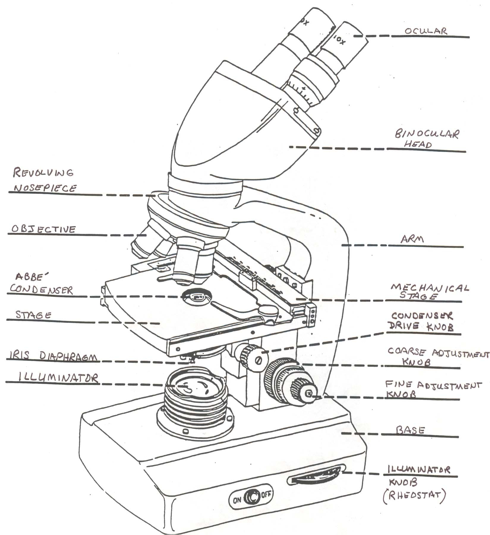
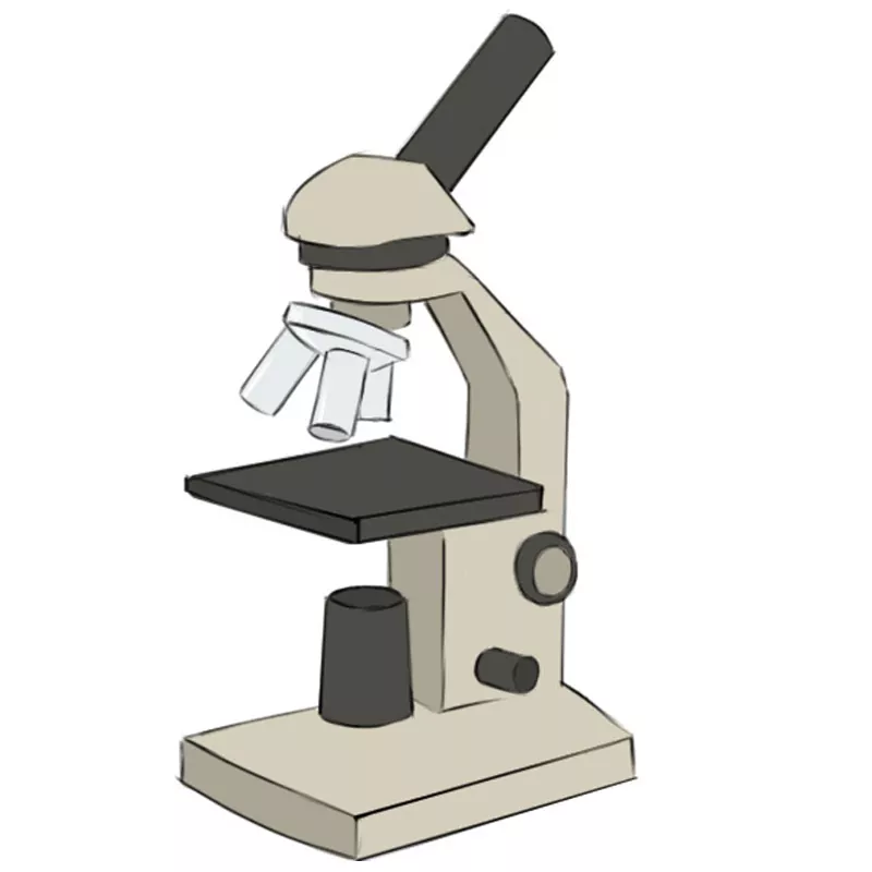
To make observations and draw scale diagrams of cells. By following the simple steps, you too can easily draw a perfect microscope. Web a light microscope is a biology laboratory instrument or tool, that uses visible light to detect and magnify very small objects and enlarge them. The specimen is normally placed close to the microscopic lens. From wikimedia commons, the free media repository. Microscopes can be simple or complex in design, and some can do more than one type of microscopy, each of which reveals slightly different information. The light transmitted from the specimen enters the objective lens. Light and electron microscopes allow us to see inside cells. Knob used to adjust the amount of light that reaches the specimen or slide from the base illumination. Stage clips hold the slide in place c.
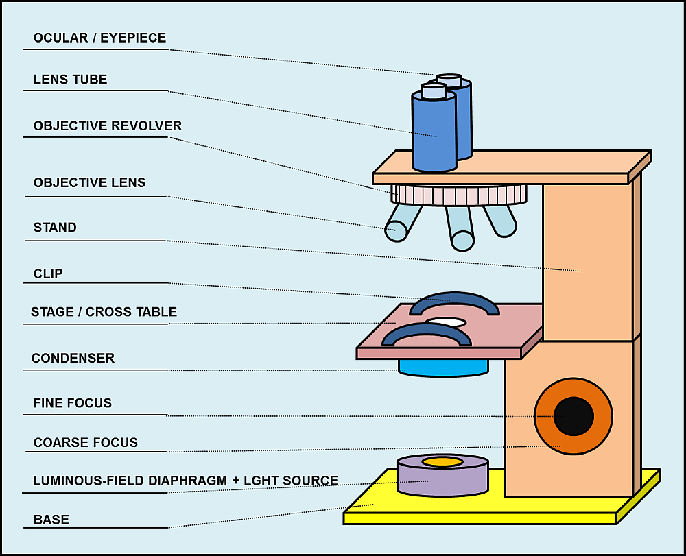
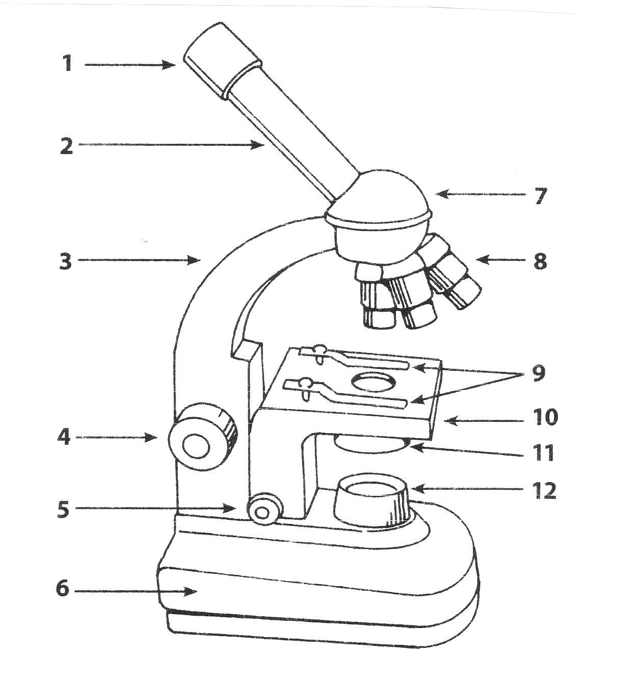
The specimen is normally placed close to the microscopic lens. Optical microscopes are the oldest design of microscope and were possibly invented in their present compound form in the 17th century. Specimens must be prepared on a microscope slide to be observed under a light microscope. The parts of a light microscope and their functions. Place the second magnifying glass between your eye and the first magnifying glass. Diaphragm regulates the amount of light on the specimen e. Web in a simple light microscope, a thin specimen containing a slide is placed on the microscope’s stage. We are going to begin our microscope drawing by creating the upper eyepiece. Objective lenses magnification ranges from 10 x to 40 x f. Stage supports the slide being.
Light Microscope Drawing at GetDrawings Free download
By following the simple steps, you too can easily draw a perfect microscope. Low voltage halogen bulbs are the most commonly used source of illumination for compound microscopes. This must be done carefully to avoid damaging any biological specimen. Bottom lens or field diaphragm: Objective lenses magnification ranges from 10 x to 40 x f.
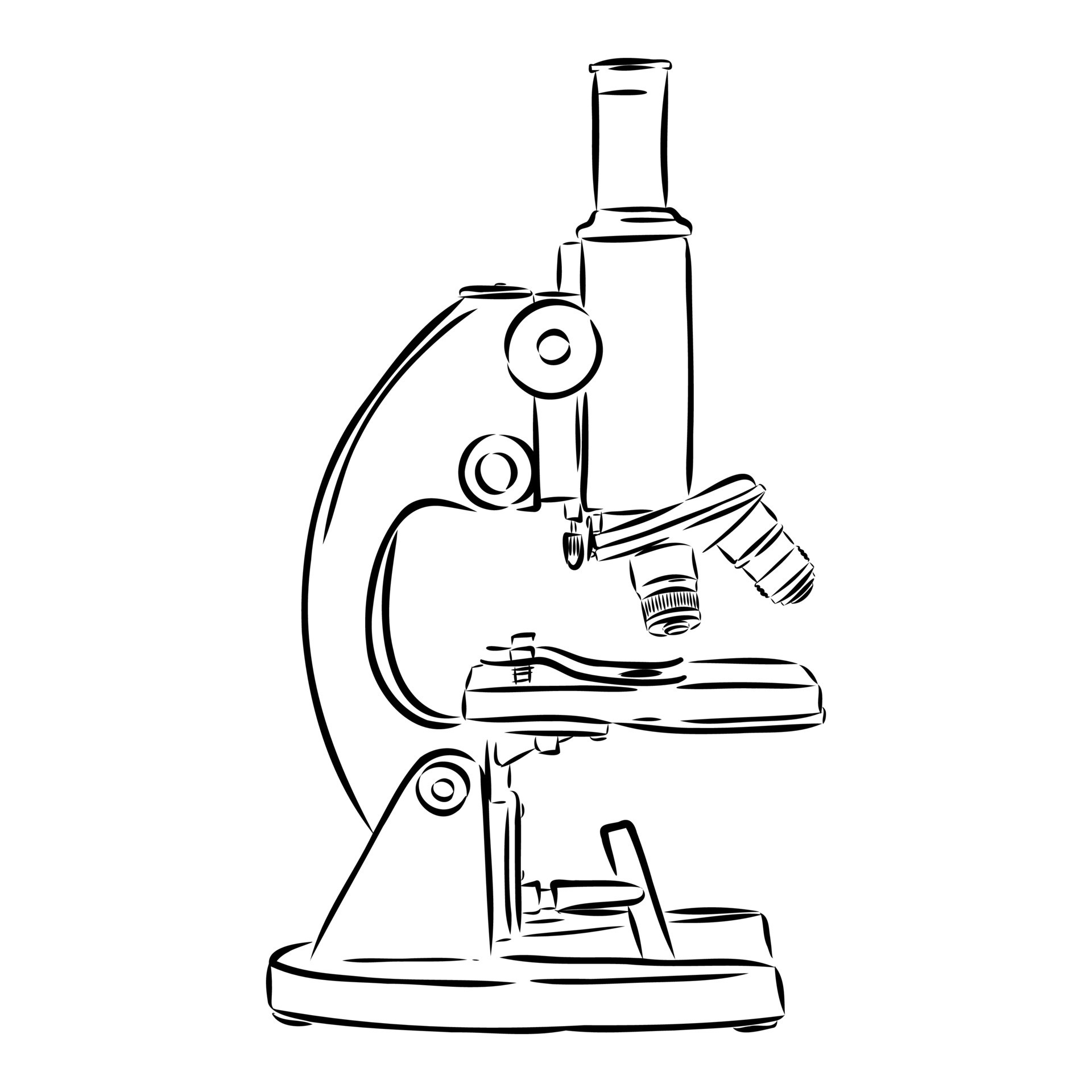
microscope vector sketch 7307564 Vector Art at Vecteezy
Here, draw a slightly angled rectangular shape. Knob used to adjust the amount of light that reaches the specimen or slide from the base illumination. Microscopes can be simple or complex in design, and some can do more than one type of microscopy, each of which reveals slightly different information. Diaphragm regulates the amount of light on the specimen e..
Light Microscope Drawing at GetDrawings Free download
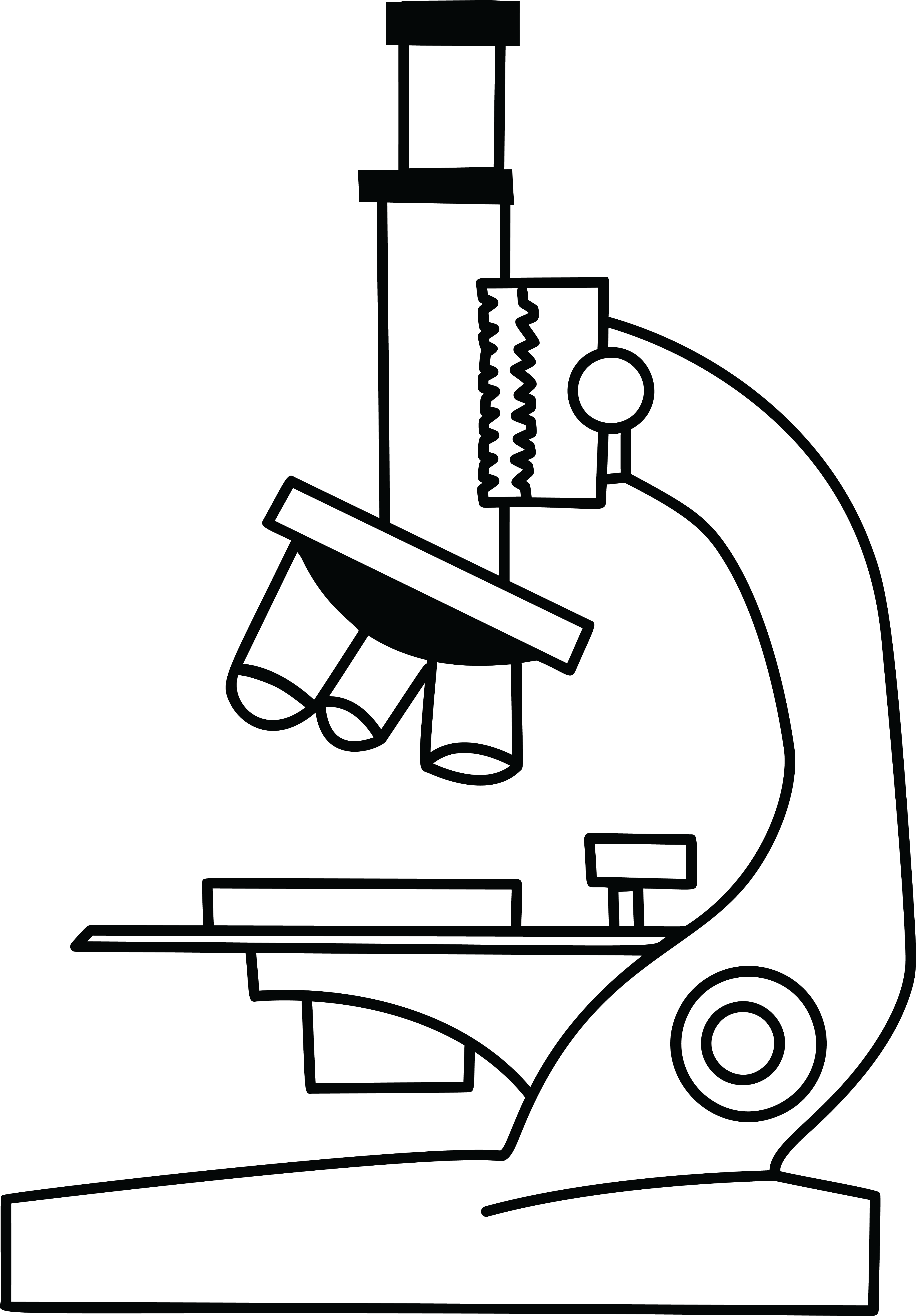
We are going to begin our microscope drawing by creating the upper eyepiece. This must be done carefully to avoid damaging any biological specimen. The light transmitted from the specimen enters the objective lens. By following the simple steps, you too can easily draw a perfect microscope. Labelled drawings to review parts and uses of the parts of a microscope.
Compound Light Microscope Drawing at Explore
Move the second glass up or down until the print comes into sharp focus. The university of waikato te whare wānanga o waikato published 8 june 2018referencing hub media. Web a light microscope is a biology laboratory instrument or tool, that uses visible light to detect and magnify very small objects and enlarge them. The light transmitted from the specimen.

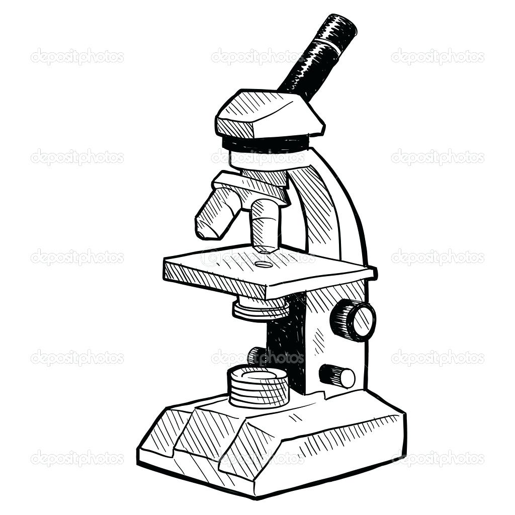
How to Draw a Microscope Really Easy Drawing Tutorial
In a light microscope, visible light passes through the specimen (the biological sample you are looking at) and is bent through the lens system, allowing the user to see a magnified image. Microscopes can be simple or complex in design, and some can do more than one type of microscopy, each of which reveals slightly different information. Use this interactive.
How to Draw a Microscope Easy Drawing Art
Objective lenses magnification ranges from 10 x to 40 x f. Bottom lens or field diaphragm: Web a video of a cell dividing to show you the power and usefulness of the microscope in science. Here, draw a slightly angled rectangular shape. Place the second magnifying glass between your eye and the first magnifying glass.
Microscope Drawing Easy at Explore collection of
Stage supports the slide being. The image of the print will look a little bit larger. From wikimedia commons, the free media repository. Stage clips hold the slide in place c. The university of waikato te whare wānanga o waikato published 8 june 2018referencing hub media.
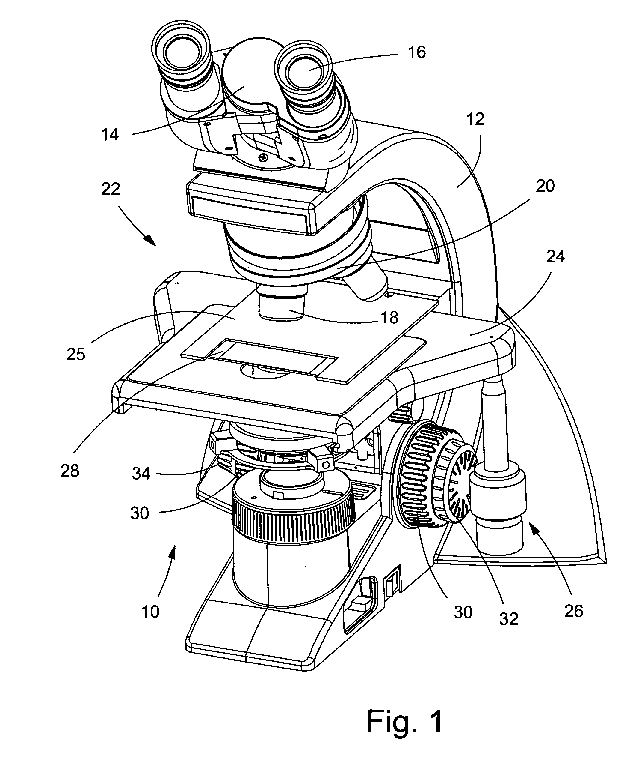
Parts and components of light microscopes Light Microscope
A light microscope can be used to observe animal and plant cells. The light transmitted from the specimen enters the objective lens. Parts of a compound microscope with diagram explained. Web light microscopy is used to make small structures and samples visible by providing a magnified image of how they interact with visible light, e.g., their absorption, reflection and scattering..
Microscope Drawing Easy at Explore collection of
By following the simple steps, you too can easily draw a perfect microscope. Web review the principles of light microscopy and identify the major parts of the microscope. Role of refraction in light microscopes. Web what is refraction? Web light microscopy is used to make small structures and samples visible by providing a magnified image of how they interact with.

Simple Microscope Definition, Principle, Magnification, Parts
Web what is refraction? Web the optical microscope, also referred to as a light microscope, is a type of microscope that commonly uses visible light and a system of lenses to generate magnified images of small objects. Low voltage halogen bulbs are the most commonly used source of illumination for compound microscopes. The light transmitted from the specimen enters the.
Objective Lenses Magnification Ranges From 10 X To 40 X F.
Plant, animal and bacterial cells have smaller components each with a specific. Begin by finding the central point of your drawing area. Use this interactive to identify and label the main parts of a microscope. Diaphragm regulates the amount of light on the specimen e.
Microscopes Can Be Simple Or Complex In Design, And Some Can Do More Than One Type Of Microscopy, Each Of Which Reveals Slightly Different Information.
Bottom lens or field diaphragm: They use lenses to focus light on the specimen, magnifying it thus producing an image. Web the optical microscope, also referred to as a light microscope, is a type of microscope that commonly uses visible light and a system of lenses to generate magnified images of small objects. Stage clips hold the slide in place c.
Specimens Must Be Prepared On A Microscope Slide To Be Observed Under A Light Microscope.
Web what is refraction? Role of refraction in light microscopes. By following the simple steps, you too can easily draw a perfect microscope. How to use a light microscope?
This Must Be Done Carefully To Avoid Damaging Any Biological Specimen.
Parts of a compound microscope with diagram explained. Light source projects light upwards through the diaphragm, the specimen, and the lenses h. Light and electron microscopes allow us to see inside cells. From wikimedia commons, the free media repository.