Loose Connective Tissue Drawing
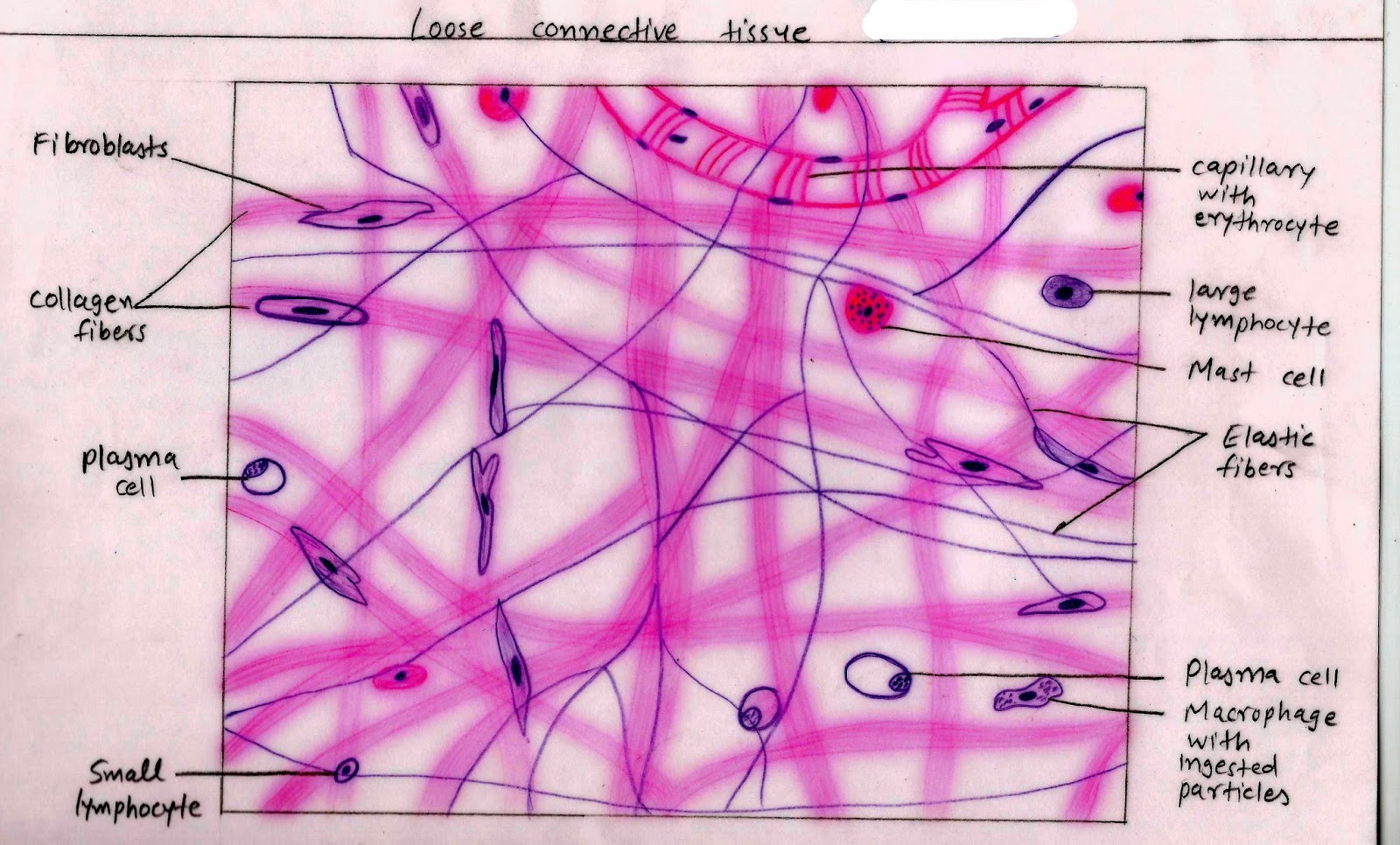
Loose Connective Tissue Drawing - Loose connective tissue is found between many organs where it acts both to absorb shock and bind tissues together. They are sketches from selected slides used in class from the. They usually stain pink and are the thickest fiber. Web #histologydrawing#histology #connectivetissuehistology#connectivetissuediagram#connectivetissue #howtodrawlooseconnectivetissue#howtodrawhistodiagram Web in loose connective tissue, the fibers are loosely organized, leaving large spaces in between. It consists of a loose irregular network of elastin fibers and collagen fibers suspended within a. This image of loose connective tissue shows collagenous fibers (red), elastic fibers (black), matrix, and fibroblasts (cells that produce the fibers). Its ground substance occupies more volume than the fibers do. Web in drawing images of connective tissue proper preparations seen under the microscope, it is important to simplify the visuals. Why do we call it “loose” connective tissue?
The drawings of histology images were originally designed to complement the histology component of the first year medical course run prior to 2004. This image of loose connective tissue shows collagenous fibers (red), elastic fibers (black), matrix, and fibroblasts (cells that produce the fibers). It's also known as areolar connective tissue, an example of which can be seen in this micrograph. The fibers and other components of the connective tissue matrix are secreted by fibroblasts. It is the predominant type of connective tissue that joins the cells in the other main tissues (muscle, nerve, and epithelia) and that joins tissues into organs. Like all tissue types, it consists of cells surrounded by a compartment of fluid called the extracellular matrix (ecm). Web loose connective tissue is a type of connective tissue proper. Connective tissue preparations are often messy with a number of blotches and shapes irrelevant to the main components of the tissue, which are the cells and the extracellular protein fibers. Web loose connective tissue is the most widely distributed of all connective tissues. Web the first pages illustrate introductory concepts for those new to microscopy as well as definitions of commonly used histology terms.
Connective tissue preparations are often messy with a number of blotches and shapes irrelevant to the main components of the tissue, which are the cells and the extracellular protein fibers. Its cellular content is highly abundant and varied. Web about press copyright contact us creators advertise developers terms privacy policy & safety how youtube works test new features nfl sunday ticket press copyright. In vertebrates, the most common type of connective tissue is loose connective tissue. Why do we call it “loose” connective tissue? Web loose connective tissue is a type of connective tissue proper. Mesentery is an example of loose (areolar) connective tissue. Web loose connective tissue proper: Web the first pages illustrate introductory concepts for those new to microscopy as well as definitions of commonly used histology terms. Loose connective tissue is composed of loosely woven collagen and elastic fibers.

Mammalian Tissues Lab Notebook Students Coursework
Why do we call it “loose” connective tissue? Web loose/areolar connective tissue. Mesentery is an example of loose (areolar) connective tissue. They usually stain pink and are the thickest fiber. Other components include collagen fibers (c) and elastic fibers (ef) read more about loose connective tissue, 40x

Loose Connective Tissue histology drawingHow to draw loose Connective
It allows water, salts, and various nutrients to diffuse through to adjacent or embedded cells and tissues. Collagen fibers are found in most supporting tissues and collagen is the most abundant protein in the body (wheaters). This chapter will focus on the types of loose connective tissue proper. Mesentery is an example of loose (areolar) connective tissue. Web loose/areolar connective.
Connective Tissue; Structure and Function McIsaac Health Systems Inc.
In vertebrates, the most common type of connective tissue is loose connective tissue. Web in loose connective tissue, the fibers are loosely organized, leaving large spaces in between. Web loose connective tissue is the most widely distributed of all connective tissues. Web about press copyright contact us creators advertise developers terms privacy policy & safety how youtube works test new.

10 b Connective tissue loose areolaradipose... Dense regular
Web loose connective tissue, also known as areolar tissue, is a cellular connective tissue with thin and relatively sparse collagen fibers. Other components include collagen fibers (c) and elastic fibers (ef) read more about loose connective tissue, 40x Its cellular content is relatively highly abundant and varied and it typically contains cells such as fibroblasts and adipocytes, fibers such as.
connective tissue anatomy and physiology
They are small purple cells found inside the connective tissue The drawings of histology images were originally designed to complement the histology component of the first year medical course run prior to 2004. Like all tissue types, it consists of cells surrounded by a compartment of fluid called the extracellular matrix (ecm). Make note of these structures and draw them.
Histology Image Connective tissue
They are small purple cells found inside the connective tissue Web in loose connective tissue, the fibers are loosely organized, leaving large spaces in between. The ecm is composed of a moderate amount of ground substance and two main types of protein fibers: Web the first pages illustrate introductory concepts for those new to microscopy as well as definitions of.
Anatomy & Physiology Connective Tissue Proper ditki medical
This chapter will focus on the types of loose connective tissue proper. Connective tissue preparations are often messy with a number of blotches and shapes irrelevant to the main components of the tissue, which are the cells and the extracellular protein fibers. Its cellular content is highly abundant and varied. Web loose/areolar connective tissue. Other components include collagen fibers (c).

Areolar Loose Connective Tissue
Web loose connective tissue with nuclei (n) labeled. Its ground substance occupies more volume than the fibers do. Web recognize different types of connective tissue (e.g., dense irregular, dense regular, loose, adipose) and know examples where they are found in the body. This image of loose connective tissue shows collagenous fibers (red), elastic fibers (black), matrix, and fibroblasts (cells that.

20 How to Draw Loose & Dense Regular Connective Tissue/Histology/1st
Web after the epithelium, i am posting videos on how to draw connective tissue. Web loose connective tissue, also known as areolar tissue, is a cellular connective tissue with thin and relatively sparse collagen fibers. They are sketches from selected slides used in class from the. Collagen fibers are found in most supporting tissues and collagen is the most abundant.

What Is Loose Connective Tissue? (preview) Human Anatomy Kenhub
A few distinct cell types and densely packed fibers in a matrix characterize these tissues. It is the predominant type of connective tissue that joins the cells in the other main tissues (muscle, nerve, and epithelia) and that joins tissues into organs. Fibroblasts are the cells that produce collagen and elastin that form the fibers found in connective tissue. Web.
The Fibers And Other Components Of The Connective Tissue Matrix Are Secreted By Fibroblasts.
Examine this section to observe collagen fibers and their fibroblasts. Web #histologydrawing#histology #connectivetissuehistology#connectivetissuediagram#connectivetissue #howtodrawlooseconnectivetissue#howtodrawhistodiagram Web the first pages illustrate introductory concepts for those new to microscopy as well as definitions of commonly used histology terms. Web loose connective tissue.
Web Loose/Areolar Connective Tissue.
Web loose connective tissue, also known as areolar tissue, is a cellular connective tissue with thin and relatively sparse collagen fibers. Connective tissue is the tissue that connects or separates, and supports all the other types of tissues in the body. Collagen fibers are found in most supporting tissues and collagen is the most abundant protein in the body (wheaters). Supportive connective tissue —bone and cartilage—provide structure and strength to the body and protect soft tissues.
Loose Connective Tissue, Also Called Areolar Connective Tissue, Has A Sampling Of All Of The Components Of A Connective Tissue.
Web loose connective tissue (lct), also called areolar tissue, belongs to the category of connective tissue proper. Its ground substance occupies more volume than the fibers do. Web after the epithelium, i am posting videos on how to draw connective tissue. Connective tissue is one of the basic tissue types of the body.
Loose Connective Tissue Is Found Between Many Organs Where It Acts Both To Absorb Shock And Bind Tissues Together.
Loose connective tissue is composed of loosely woven collagen and elastic fibers. They are sketches from selected slides used in class from the. They are small purple cells found inside the connective tissue Web in drawing images of connective tissue proper preparations seen under the microscope, it is important to simplify the visuals.