Lumbar Plexus Drawing
Lumbar Plexus Drawing - Web the lumbar plexus is a network of nerve fibres that supplies the skin and musculature of the lower limb. Web how to draw the lumbosacral plexus. Web about press copyright contact us creators advertise developers terms privacy policy & safety how youtube works test new features nfl sunday ticket press copyright. Discuss key mri sequences and present an mri sequence‐based search pattern. Web the lumbosacral plexus is formed by the anterior rami (i.e., branches) of spinal nerves l4 to l5 and s1 to s4. 330k views 12 years ago. The sacral plexus is located on the posterior pelvic wall, posterior to the internal iliac vessels and ureter, and anterior to the piriformis muscle. Triangular form, with its base resting against the lumbar vertebrae and. The anterior roots from l1 to l3 and the greater part of l4. I decided that i would combine what i.
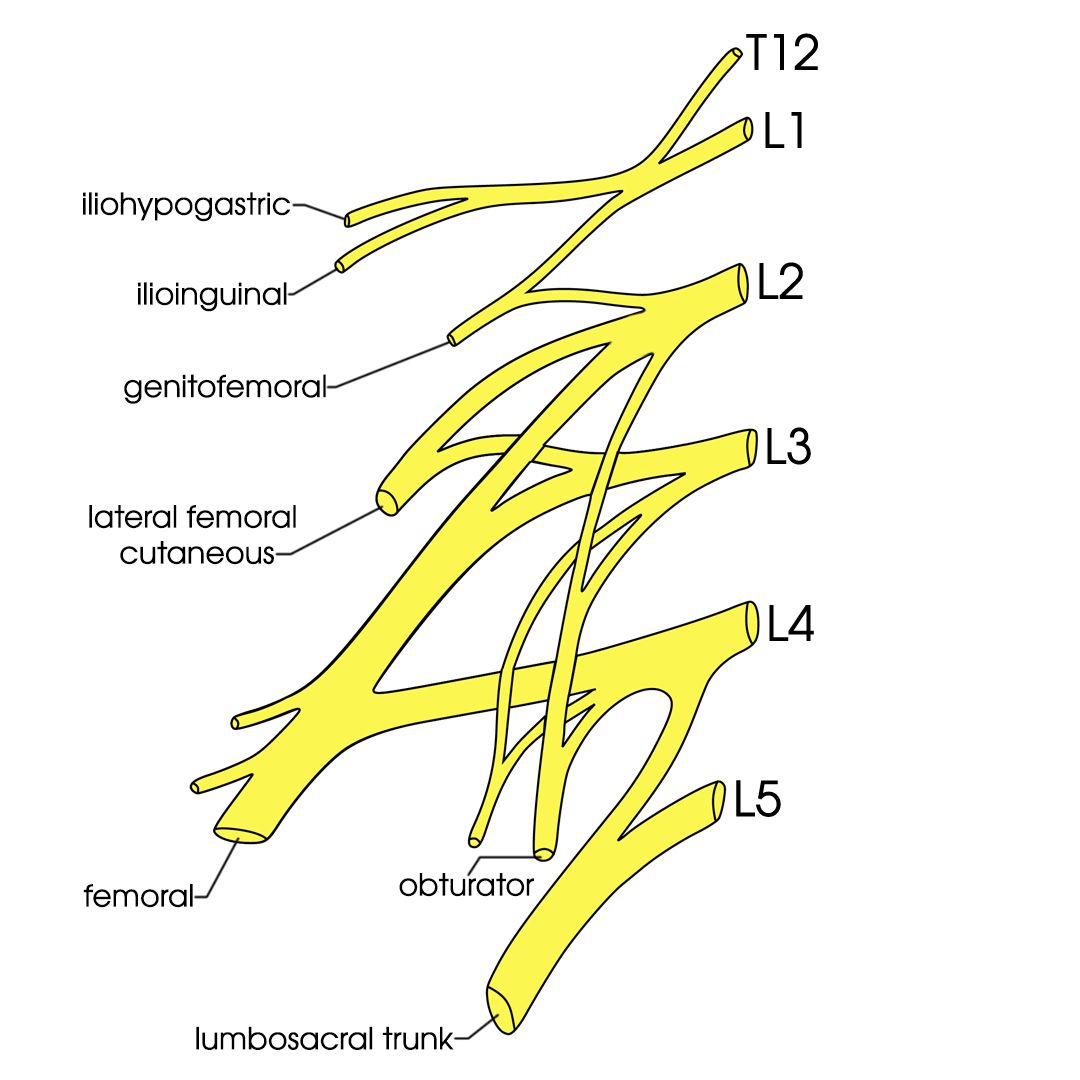
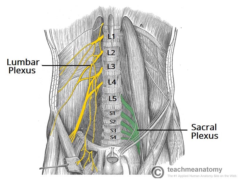
It is located on the posterolateral wall of the lesser pelvis, adjacent to the lumbar spine. Web the lumbosacral plexus is formed by the anterior rami (i.e., branches) of spinal nerves l4 to l5 and s1 to s4. Web how to draw the lumbosacral plexus. The anterior roots from l1 to l3 and the greater part of l4. It is located on the posterior abdominal wall, anterior to the transverse processes of the lumbar vertebrae and within the posterior portion of the psoas major muscle. Discuss role of mr imaging of the lumbosacral plexus in clinical practice. Veretrebral transverse process and its apex formed by the union of the. They are formed where t12 to l5 exit the spinal cord via intervertebral foramina. The lumbar plexus is an essential collection of nerves that arise from mostly the lumbar spinal cord. Note the connections between these nerves and the more anteriorly placed sympathetic trunk, which then forms an extensive plexus over the anterior longitudinal ligament shaded blue here.
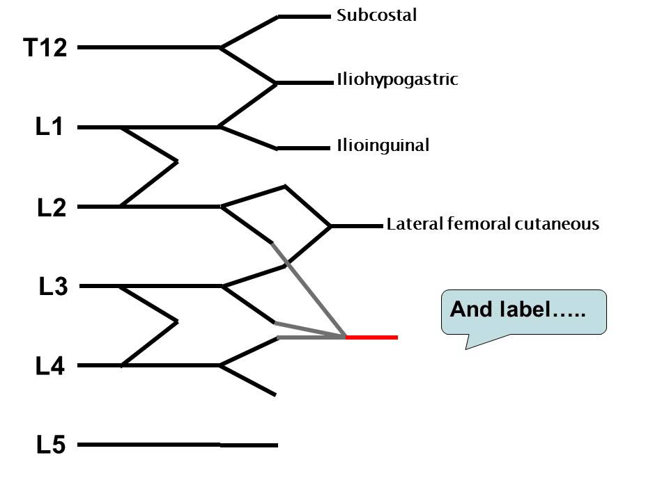
Case courtesy of assoc prof craig hacking, radiopaedia.org. The sacral plexus is located on the posterior pelvic wall, posterior to the internal iliac vessels and ureter, and anterior to the piriformis muscle. When i was taking medical gross anatomy, i couldn't find a good video that included the lumbar and sacral plexus. The anterior roots from l1 to l3 and the greater part of l4. Third roots with the ascendant rami of the fourth. Ventral rami of t12 to l5. The lumbar plexus is formed from the anterior rami of l1 to l4. The structure of the lumbar plexus including its spinal roots and branches, which supply the abdominal wall, pelvis and lower limb. Web drawing the lumbosacral plexus and learning from which spinal segments do the nerves originate. Web about press copyright contact us creators advertise developers terms privacy policy & safety how youtube works test new features nfl sunday ticket press copyright.

Lumbar plexus clinical anatomy spinal nerves Vector Image
When i was taking medical gross anatomy, i couldn't find a good video that included the lumbar and sacral plexus. Web how to draw the lumbosacral plexus. Review lumbosacral plexus normal anatomy and important normal variant anatomy. Web the lumbar plexus is a network of nerve fibres that supplies the skin and musculature of the lower limb. The lumbar plexus.
How to draw the lumbar plexus YouTube
We’re going to look at the basic structure and the location of the lumbar plexus and a few ways to remember some of the nerves and the basic organization. 330k views 12 years ago. Draw it twice and am sure you will be. It lies within the psoas major muscle and gives rise to several important nerves, including: The sacral.
Lumbar plexus Anatomy, branches and innervation Kenhub
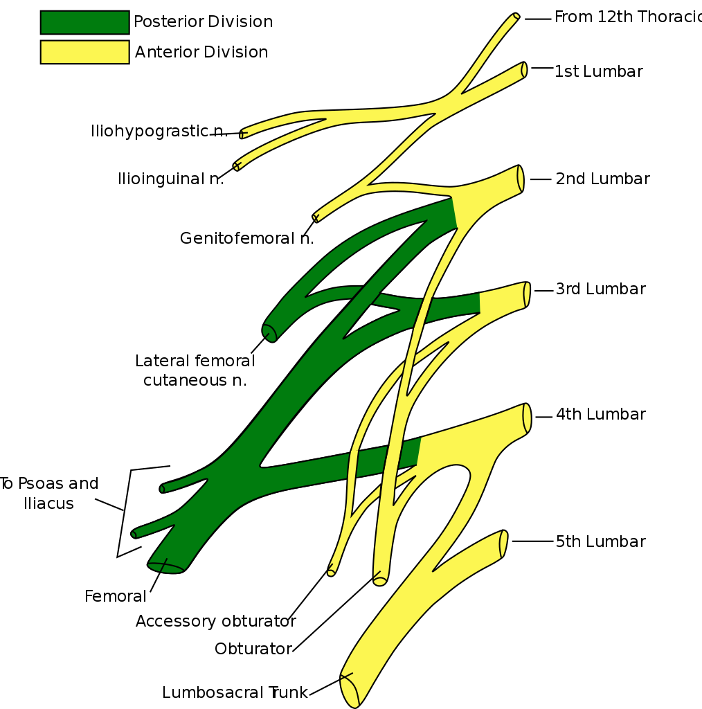
It lies within the psoas major muscle and gives rise to several important nerves, including: We’re going to look at the basic structure and the location of the lumbar plexus and a few ways to remember some of the nerves and the basic organization. The lumbosacral plexus then embeds itself into the psoas major muscle and later emerges in the.
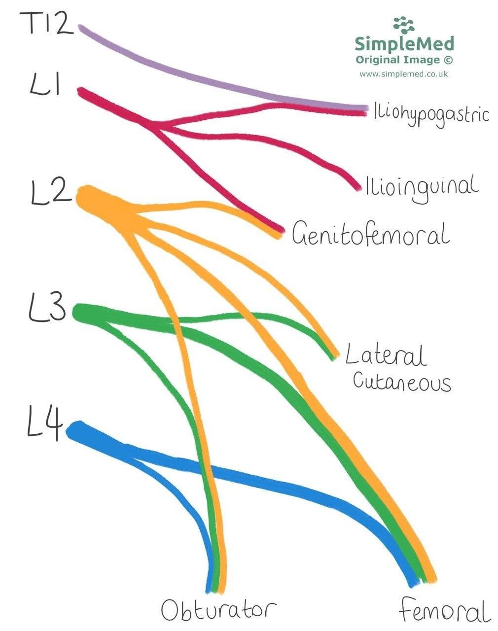
11. The Lumbar Plexus SimpleMed Learning Medicine, Simplified
Web how to draw the lumbosacral plexus. Case courtesy of assoc prof craig hacking, radiopaedia.org. Web the lumbosacral plexus is formed by the anterior rami (i.e., branches) of spinal nerves l4 to l5 and s1 to s4. The lumbosacral plexus then embeds itself into the psoas major muscle and later emerges in the pelvis. It is located in the lumbar.

Schematic drawing of lumbar and sacral plexus and the main pelvic
Web the lumbar plexus is a network of nerve fibres that supplies the skin and musculature of the lower limb. The lumbosacral plexus then embeds itself into the psoas major muscle and later emerges in the pelvis. Draw it twice and am sure you will be. It is located on the posterolateral wall of the lesser pelvis, adjacent to the.

Lumbosacral Plexus What Is It, Nerves, and More Osmosis
Web drawing the lumbar plexus is a very easy process once you get the muscle memory down. Third roots with the ascendant rami of the fourth. Discuss role of mr imaging of the lumbosacral plexus in clinical practice. Web the lumbar plexus is a network of nerve fibres that supplies the skin and musculature of the lower limb. Web about.
Lumbosacral Plexus & Lower Extremity Neuropathies Exam Review
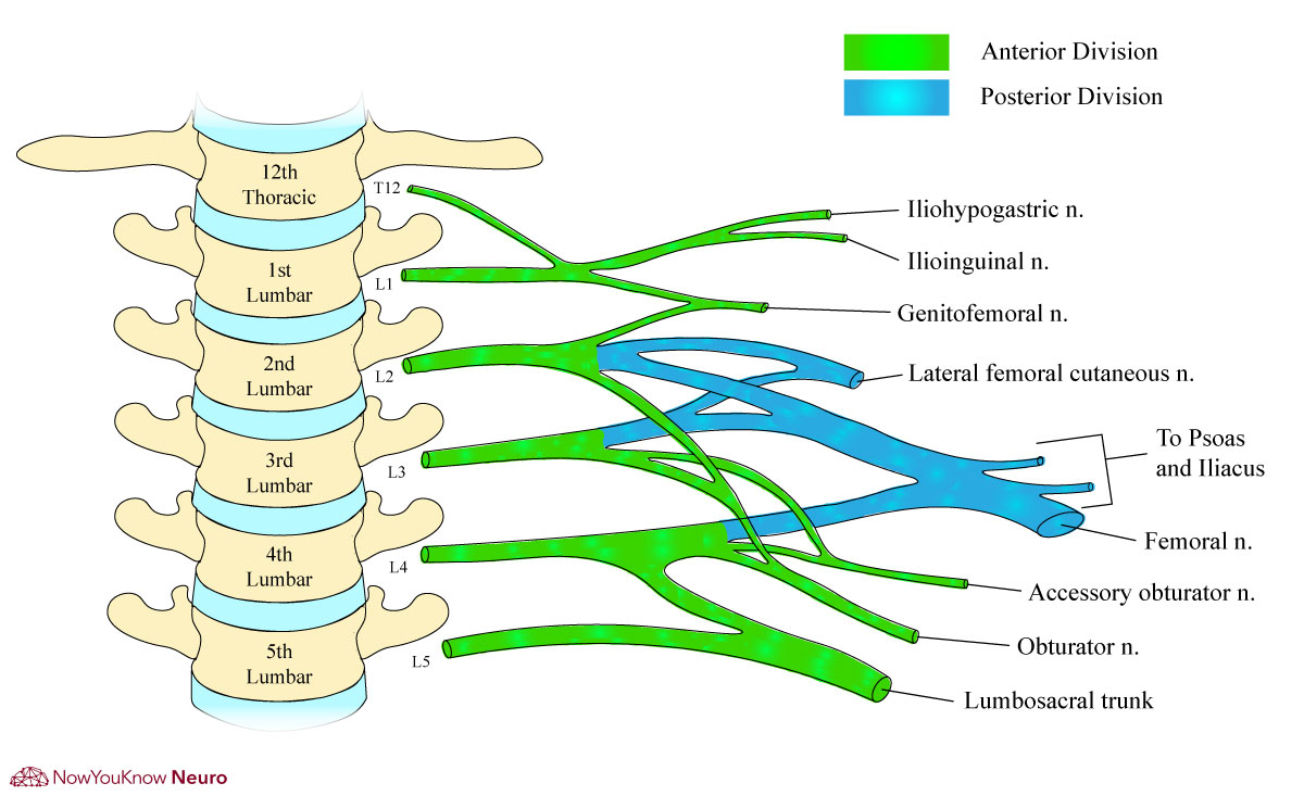
Web the lumbar plexus is a complex neural network formed by the lower thoracic and lumbar ventral nerve roots (t12 to l5) which supplies motor and sensory innervation to the lower limb and pelvic girdle. It is located on the posterior abdominal wall, anterior to the transverse processes of the lumbar vertebrae and within the posterior portion of the psoas.
Lumbar Plexus AnatomyZone
We’re going to look at the basic structure and the location of the lumbar plexus and a few ways to remember some of the nerves and the basic organization. With the lumbar plexus drawing you can determine the nerves and the innervations. Web the lumbar plexus is a complex neural network formed by the lower thoracic and lumbar ventral nerve.
The Lumbar Plexus Spinal Nerves Branches TeachMeAnatomy
Web about press copyright contact us creators advertise developers terms privacy policy & safety how youtube works test new features nfl sunday ticket press copyright. Discuss role of mr imaging of the lumbosacral plexus in clinical practice. Its very easy once you know the scheme of lines. It is located in the lumbar region, within the substance of the psoas.
Lumbar plexus
The lumbosacral plexus then embeds itself into the psoas major muscle and later emerges in the pelvis. Web drawing the lumbar plexus is a very easy process once you get the muscle memory down. It is located on the posterior abdominal wall, anterior to the transverse processes of the lumbar vertebrae and within the posterior portion of the psoas major.
It Also Receives Some Fibers From Thoracic Nerve, T12.
Discuss role of mr imaging of the lumbosacral plexus in clinical practice. Review the mri appearance of normal and abnormal peripheral nerves. It is located on the posterolateral wall of the lesser pelvis, adjacent to the lumbar spine. The lumbar plexus is an essential collection of nerves that arise from mostly the lumbar spinal cord.
The Lumbar Plexus Is A Complex Neural Network Formed By The Lower Thoracic And Lumbar Ventral Nerve Roots.
Web the lumbosacral plexus is formed by the anterior rami (i.e., branches) of spinal nerves l4 to l5 and s1 to s4. The structure of the lumbar plexus including its spinal roots and branches, which supply the abdominal wall, pelvis and lower limb. Discuss key mri sequences and present an mri sequence‐based search pattern. It is located on the posterior abdominal wall, anterior to the transverse processes of the lumbar vertebrae and within the posterior portion of the psoas major muscle.
Third Roots With The Ascendant Rami Of The Fourth.
The sacral plexus is located on the posterior pelvic wall, posterior to the internal iliac vessels and ureter, and anterior to the piriformis muscle. Its very easy once you know the scheme of lines. This is a brief tutorial on the lumbar plexus. Veretrebral transverse process and its apex formed by the union of the.
We’re Going To Look At The Basic Structure And The Location Of The Lumbar Plexus And A Few Ways To Remember Some Of The Nerves And The Basic Organization.
I decided that i would combine what i. It lies within the psoas major muscle and gives rise to several important nerves, including: The lumbar plexus is an anastomotic complex formed by. Ventral rami of t12 to l5.