Microscope Drawing With Label
Microscope Drawing With Label - Microscope, which were created in the 16th century and contain the ability to enlarge microscopic objects like microbial cells, have revolutionised science by producing images containing recognisable as well as distinguishable. Web use this interactive to identify and label the main parts of a microscope. 197k views 3 years ago how to draw back to school! The microscopes parts divided into three different structural parts head, base, and arms. A microscope is a laboratory instrument used to examine objects that are too small to be seen by the naked eye. In this tutorial, writing master shows you how to draw. Coarse and fine focus knobs. There are three structural parts of the microscope i.e. Web we have explained how to draw a microscope step by step easy and realistic in this article. 📏 the microscope has three major structural parts:
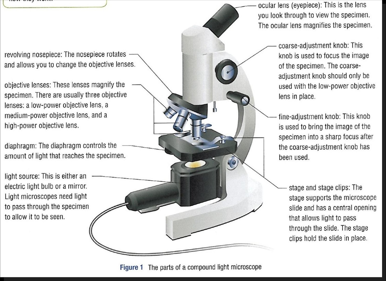
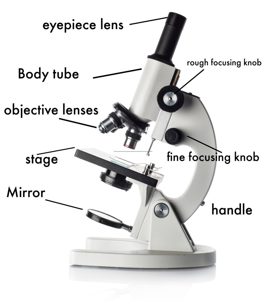
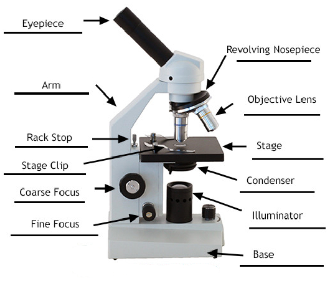
Eyepiece (ocular lens) with or without pointer: 📏 the microscope has three major structural parts: The eyepiece usually contains a 10x or 15x power lens. Web labeled diagram of a compound microscope. Drag and drop the text labels onto the microscope diagram. 👩🎨 join our art hub membership! 800.942.0528 (us toll free) 1.760.438.0528 (international) microscope world explains the parts of the microscope, including a printable worksheet for schools and home. Parts of a microscope with functions and labeled diagram. A microscope is a laboratory instrument used to examine objects that are too small to be seen by the naked eye. Web 🌟 there are several important parts of the microscope that contribute to its proper functioning, including the eyepiece, objective lenses, focus knobs, stage, light source, and condenser.
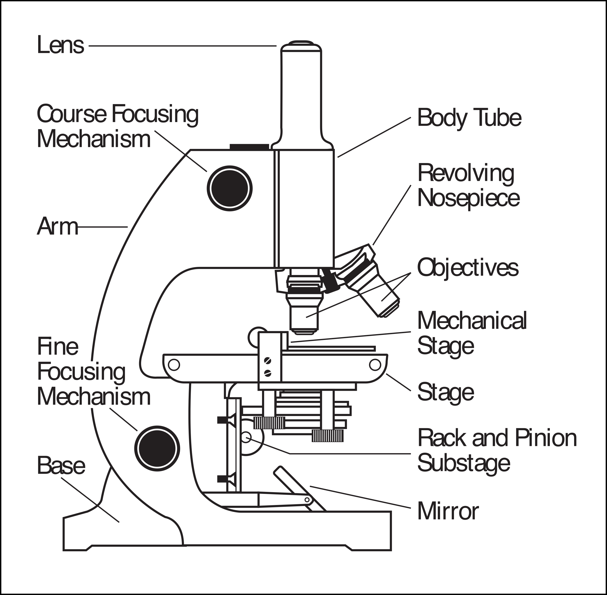
The microscopes parts divided into three different structural parts head, base, and arms. There is a blank copy at the end of the video to review. There are three structural parts of the microscope i.e. Web learn about the different parts of the microscope, including the simple microscope and the compound microscope, with labeled pictures and detailed explanations. In this tutorial, writing master shows you how to draw. Web with these steps, you've mastered how to draw a microscope and created a realistic looking microscope picture. Microscopes are useful instruments in a variety of sectors, ranging from scientific inquiry to medical diagnostics. Major structural parts of a compound microscope. The eyepiece usually contains a 10x or 15x power lens. Coarse and fine focus knobs.

301 Moved Permanently
800.942.0528 (us toll free) 1.760.438.0528 (international) microscope world explains the parts of the microscope, including a printable worksheet for schools and home. The optical parts of a simple microscope include. First and foremost, we have a labeled microscope diagram, available in both black and white and color. The eyepiece usually contains a 10x or 15x power lens. 1k views 4.

Parts of a Microscope SmartSchool Systems
First and foremost, we have a labeled microscope diagram, available in both black and white and color. Structural support that holds & connects the eyepieces to the objective lenses. Use this with the microscope parts activity to help students identify and label the main parts of a microscope and then describe their functions. Eyepiece (ocular lens) with or without pointer:.

Compound Microscope Parts, Functions, and Labeled Diagram New York
The lens the viewer looks through to see the specimen. You can add labels to the various components for educational purposes or leave it as it is. 📐 adjustment knobs are used to adjust the focus of the microscope. Web learn about the different parts of the microscope, including the simple microscope and the compound microscope, with labeled pictures and.

Parts of a microscope with functions and labeled diagram (2023)
We have many other exciting drawing tutorials that you can try, and we hope to see you soon! Drag and drop the text labels onto the microscope diagram. Today, we're learning how to draw a cool microscope! The eyepiece usually contains a 10x or 15x power lens. Web labeled diagram of simple microscope parts.
Parts Of Microscope
Label the parts of the microscope with answers (a4) pdf print version. 800.942.0528 (us toll free) 1.760.438.0528 (international) microscope world explains the parts of the microscope, including a printable worksheet for schools and home. Ready to take your drawing skills to the next level? You can add labels to the various components for educational purposes or leave it as it.
Clipart microscope parts labeled WikiClipArt
Major structural parts of a compound microscope. 📏 the microscope has three major structural parts: In other words, it enlarges images of small objects. Web labeled diagram of a compound microscope. Web labeled diagram of simple microscope parts.
How to Use a Microscope
The head, the base, and the arm. Optical components of a compound microscope. In this video i go over a microscope drawing that is easy with label. What colors do you need for a microscope drawing? Label the parts of the microscope (a4) pdf print version.

Simple Microscope Definition, Principle, Magnification, Parts
Answers pdf printable version here. Web the description given below summarize the brief description of microscope parts used to visualize the microscopic specimens such as animal cells, plant cells, microbes, bacteria, viruses, microorganisms etc. Web download the label the parts of the microscope: Drag and drop the text labels onto the microscope diagram. Structural support that holds & connects the.
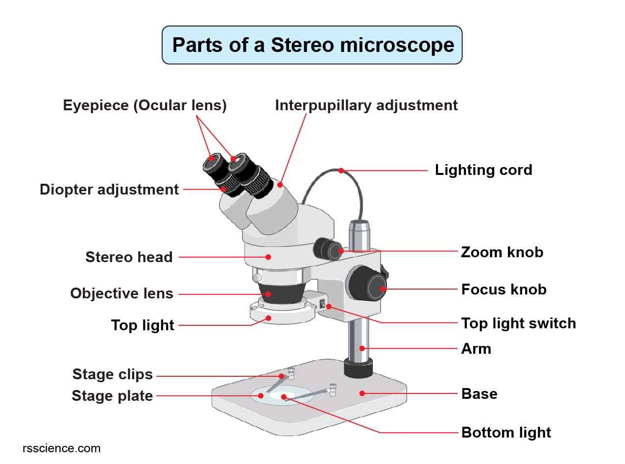
Parts of Stereo Microscope (Dissecting microscope) labeled diagram
There are three structural parts of the microscope i.e. 453 views 3 years ago #labels #chatgpt #drawing. Web the description given below summarize the brief description of microscope parts used to visualize the microscopic specimens such as animal cells, plant cells, microbes, bacteria, viruses, microorganisms etc. By raeditor / june 20, 2022. Choose from three different methods according to your.
Parts of a Compound Microscope Labeled (with diagrams) Medical
Web labeled diagram of a compound microscope. You can add labels to the various components for educational purposes or leave it as it is. Today, we're learning how to draw a cool microscope! What colors do you need for a microscope drawing? Useful as a means to change focus on one eyepiece so as to correct for any difference in.
In This Tutorial, Writing Master Shows You How To Draw.
Web with these steps, you've mastered how to draw a microscope and created a realistic looking microscope picture. Ready to take your drawing skills to the next level? The optical parts of a simple microscope include. What colors do you need for a microscope drawing?
Use This With The Microscope Parts Activity To Help Students Identify And Label The Main Parts Of A Microscope And Then Describe Their Functions.
First and foremost, we have a labeled microscope diagram, available in both black and white and color. Web we have explained how to draw a microscope step by step easy and realistic in this article. April 7, 2021 by biocheminsider. 453 views 3 years ago #labels #chatgpt #drawing.
By Raeditor / June 20, 2022.
Microscopes are useful instruments in a variety of sectors, ranging from scientific inquiry to medical diagnostics. Structural support that holds & connects the eyepieces to the objective lenses. Useful as a means to change focus on one eyepiece so as to correct for any difference in vision between your two eyes. The microscopes parts divided into three different structural parts head, base, and arms.
In This Video I Go Over A Microscope Drawing That Is Easy With Label.
800.942.0528 (us toll free) 1.760.438.0528 (international) microscope world explains the parts of the microscope, including a printable worksheet for schools and home. Microscope, which were created in the 16th century and contain the ability to enlarge microscopic objects like microbial cells, have revolutionised science by producing images containing recognisable as well as distinguishable. The part that is looked through at the top of the compound microscope. Major structural parts of a compound microscope.