Microtubules Drawing
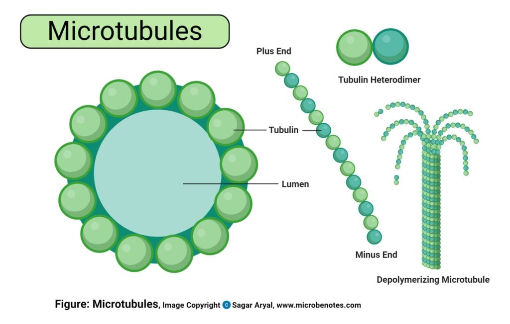
Microtubules Drawing - Each microtubule subunit comprises two closely related polypeptides: Web more specifically, in the first part of anaphase — sometimes called anaphase a — the kinetochore microtubules shorten and draw the chromosomes toward the spindle poles. Microtubules have many features that distinguish them from microfilaments and intermediate filaments. Web despite the “micro” in their name, microtubules are the largest of the three types of cytoskeletal fibers, with a diameter of about 25 nm. 1.5k views 2 years ago scientific illustration | adobe illustrator. 32k views 3 years ago. Microtubules are hollow cylinders [1] that are approximately 25nm in diameter [2] and vary in length from 200 nm to 25 μm. Microtubules, composed of alpha and beta tubulin, dynamically change length to fulfill their functions. Microtubules are essential, multitasking protein polymers that serve as structural elements in most eukaryotic cells. Microtubules are usually discussed with microfilaments.
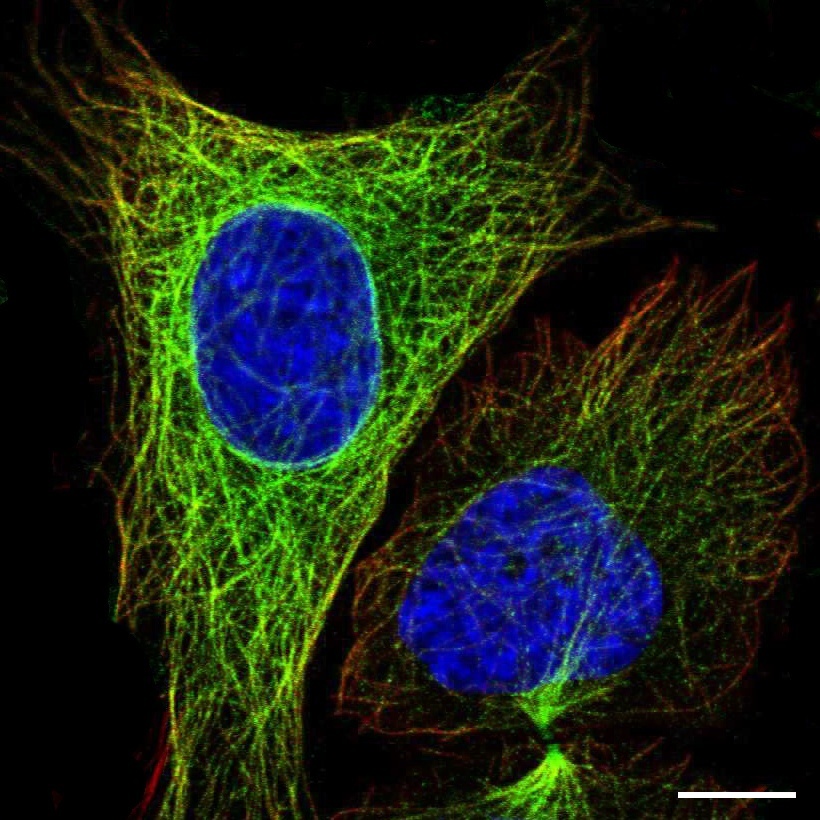
The microtubule polymer is largely viewed as a passive building block during the organization process. Web microtubules are made up of two equally distributed, structurally similar, globular subunits: Web microtubules and microfilaments have dual functions, dynamically maintaining cell shape and. Tubulin dimers can depolymerize as well as polymerize, and microtubules can undergo rapid cycles of assembly and disassembly. Web more specifically, in the first part of anaphase — sometimes called anaphase a — the kinetochore microtubules shorten and draw the chromosomes toward the spindle poles. Web dynamic networks of protein filaments give shape to cells and power cell movement. Multiple units of these dimers polymerize to form a chain called the protofilament. Microtubules, composed of alpha and beta tubulin, dynamically change length to fulfill their functions. Microtubules are essential, multitasking protein polymers that serve as structural elements in most eukaryotic cells. Web anatomy of the mitotic spindle.
To begin with, the outside diameter of a microtubule (usually about 25 nm) is much greater than that of microfilaments. Kinesin and dynein, the prototypes of microtubule motor proteins, move along microtubules in opposite directions— kinesin toward the plus end and dynein toward the minus end ( figure 11.45 ). Web anatomy of the mitotic spindle. They are dynamic, and their dynamics. They are the largest structures in the cytoskeleton and are about 24 nm thick. Biology for majors i (lumen) 6: The microtubules are cytoplasmic tubules that serve as structural components of cytoskeleton, cilia, and eukaryotic flagella. Furthermore, microtubules are hollow, containing a central lumen about 15 nm in diameter. Microtubules are the largest structures in the cytoskeleton at about 24 nanometers thick. Web microtubules are polymers of tubulin that form part of the cytoskeleton and provide structure and shape to eukaryotic cells.

Microtubules, artwork Stock Image C029/7073 Science Photo Library
Learn how microtubules, actin filaments, and intermediate filaments organize the cell. Web article 27 february 2020. Diagram indicating kinetochore microtubules (bound to kinetochores) and the aster. Microtubules are the largest structures in the cytoskeleton at about 24 nanometers thick. 32k views 3 years ago.

Structure of a microtubule, illustration Stock Image F020/1416
Like microfilaments, microtubules are also dependent on a nucleotide triphosphate for polymerization, but in this case, it is gtp. Web dynamic networks of protein filaments give shape to cells and power cell movement. Multiple units of these dimers polymerize to form a chain called the protofilament. Microtubules can be as long as 50 micrometres, as wide as 23 to 27.

Microtubules In A Cell Diagram
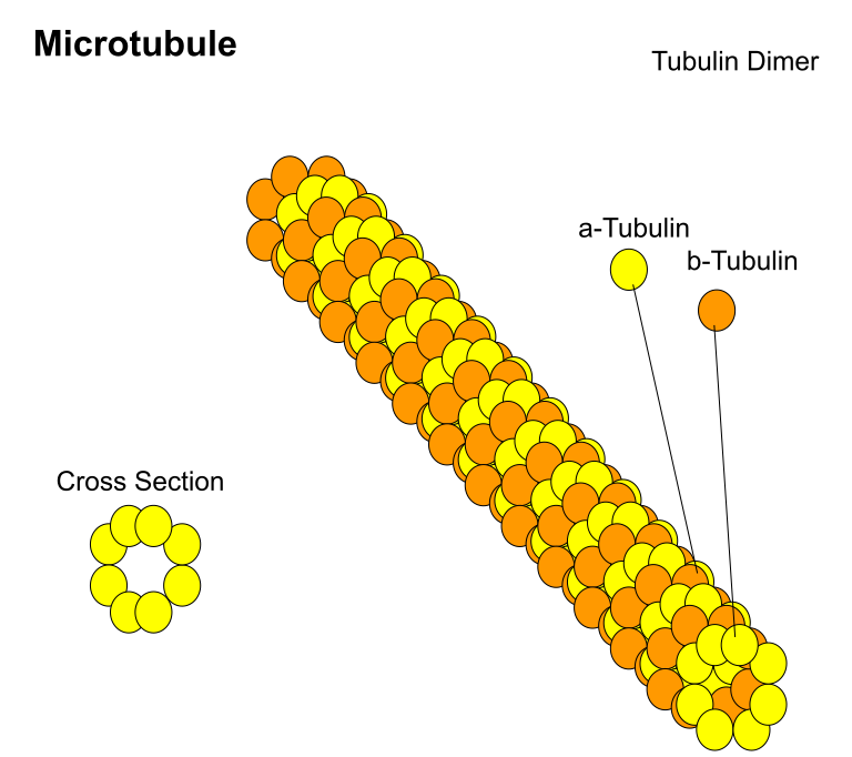
Microtubules, composed of alpha and beta tubulin, dynamically change length to fulfill their functions. Web microtubules are made up of two equally distributed, structurally similar, globular subunits: Web microtubules and microfilaments have dual functions, dynamically maintaining cell shape and. The microtubule polymer is largely viewed as a passive building block during the organization process. Microtubules have many features that distinguish.

Microtubules, illustration Stock Image F020/1405 Science Photo
Microtubules, composed of alpha and beta tubulin, dynamically change length to fulfill their functions. Microtubules are essential, multitasking protein polymers that serve as structural elements in most eukaryotic cells. To begin with, the outside diameter of a microtubule (usually about 25 nm) is much greater than that of microfilaments. Tubulin dimers can depolymerize as well as polymerize, and microtubules can.

Elements of the microtubule cytoskeleton within the cilium. Download
Web article 27 february 2020. Multiple units of these dimers polymerize to form a chain called the protofilament. As their name implies, microtubules are small hollow tubes. Kinesin and dynein, the prototypes of microtubule motor proteins, move along microtubules in opposite directions— kinesin toward the plus end and dynein toward the minus end ( figure 11.45 ). They form a.

Microtubuli Il citoscheletro BioPills
Microtubules are usually discussed with microfilaments. Diagram indicating kinetochore microtubules (bound to kinetochores) and the aster. Web dynamic networks of protein filaments give shape to cells and power cell movement. As their name implies, microtubules are small hollow tubes. This tutorial demonstrates how to draw microtubule for research publication, conference.
Cell Organelles Definition, Structure, Functions, Diagram
While microfilaments are thin, microtubules are thick, strong spirals of thousands of subunits. Biology for majors i (lumen) 6: Multiple units of these dimers polymerize to form a chain called the protofilament. Web despite the “micro” in their name, microtubules are the largest of the three types of cytoskeletal fibers, with a diameter of about 25 nm. Microtubules are essential,.
Dictionary Cell Microtubules The Human Protein Atlas
Web despite the “micro” in their name, microtubules are the largest of the three types of cytoskeletal fibers, with a diameter of about 25 nm. This tutorial demonstrates how to draw microtubule for research publication, conference. They form a network within neurons for internal transport. Each microtubule subunit comprises two closely related polypeptides: While microfilaments are thin, microtubules are thick,.
Microtubules are constituents ofA) Centrosome, nucleosome, and
Web microtubules and microfilaments have dual functions, dynamically maintaining cell shape and. Microtubules are the proteins of the cytoskeleton. Web microtubules are polymers of tubulin that form part of the cytoskeleton and provide structure and shape to eukaryotic cells. They are the largest structures in the cytoskeleton and are about 24 nm thick. Learn how microtubules, actin filaments, and intermediate.

Microtubules Biochemistry Medbullets Step 1
Multiple units of these dimers polymerize to form a chain called the protofilament. Tubulin dimers can depolymerize as well as polymerize, and microtubules can undergo rapid cycles of assembly and disassembly. They form a network within neurons for internal transport. To begin with, the outside diameter of a microtubule (usually about 25 nm) is much greater than that of microfilaments..
Furthermore, Microtubules Are Hollow, Containing A Central Lumen About 15 Nm In Diameter.
Microtubules are the proteins of the cytoskeleton. Identification of microtubule motor proteins. They are the largest structures in the cytoskeleton and are about 24 nm thick. They are dynamic, and their dynamics.
Web Despite The “Micro” In Their Name, Microtubules Are The Largest Of The Three Types Of Cytoskeletal Fibers, With A Diameter Of About 25 Nm.
Each microtubule subunit comprises two closely related polypeptides: Microtubules are hollow cylinders [1] that are approximately 25nm in diameter [2] and vary in length from 200 nm to 25 μm. Web microtubules and microfilaments have dual functions, dynamically maintaining cell shape and. Kinesin and dynein, the prototypes of microtubule motor proteins, move along microtubules in opposite directions— kinesin toward the plus end and dynein toward the minus end ( figure 11.45 ).
As Their Name Implies, Microtubules Are Small Hollow Tubes.
Then, 13 protofilaments arrange into a cylindrical pattern to form a microtubule. 1.5k views 2 years ago scientific illustration | adobe illustrator. Microtubules are essential, multitasking protein polymers that serve as structural elements in most eukaryotic cells. Web more specifically, in the first part of anaphase — sometimes called anaphase a — the kinetochore microtubules shorten and draw the chromosomes toward the spindle poles.
Of The Three Main Cytoskeletal Fibers, Intermediate Filaments Serve A Mainly Structural Role In Cells.
Microtubules are usually discussed with microfilaments. Learn how microtubules, actin filaments, and intermediate filaments organize the cell. Tubulin dimers can depolymerize as well as polymerize, and microtubules can undergo rapid cycles of assembly and disassembly. This tutorial demonstrates how to draw microtubule for research publication, conference.