Retinal Drawing
Retinal Drawing - | find, read and cite all the research you need. Web by taking the time to sketch the retinal details, residents can develop a better understanding of the anatomy, pathology, and clinical relevance of retinal findings, which can be invaluable in their training and future practice. Slit lamp biomicroscopy, smartphone funduscopy, and retinal image drawing. Published in the permanente journal 2011. Luann dvorak, phd, lpn stephen r russell, md. Web retinal drawings are a valuable tool allowing for easy visual communication of ophthalmoscopic findings despite less frequent use today in the era of digital medical records and abundance of imaging options (schachat et al. Web meet codes 92201 and 92202. Web a detailed retinal drawing must be present in the patient’s chart. Web throughout movies and simple scrolling, the ipad pro’s promotion screen uses refresh rates of up to 120hz to make sure visuals stay as smooth as its overall performance. 91 views 1 year ago.
The surface pro 9 and. First, trace the optic nerve on the retinal drawing. Without this documentation, eo is not billable. I (sr) was one of the last retina fellows in a line of… expand. Chapters will highlight differences among various artists' representations of similar diagnoses, and how drawing style and technique evolved over time, how shading—sometimes. Web most retinal surgeons are trained to create formal retinal drawings of the fundus. A lost art of medicine | find, read and cite all the research you need on researchgate. With retinal drawing and scleral depression of peripheral retinal disease (e.g., for retinal tear, retinal detachment, retinal tumor) with interpretation and report, unilateral or bilateral. Can be used for serial follow up of patients to document changes in the pathology. The two replacement codes are defined as follows:
The drawing’s size is usually a minimum of three to four inches in diameter. The lost art of retinal drawing (in progress), which will feature over 120 drawings and a history of the practice and process. Lets take a look into the most important colors for retinal drawing. Can be used for serial follow up of patients to document changes in the pathology. Acquired immunodeficiency syndrome (aids), cidofovir, cytomegalovirus (cmv), cmv retinitis fomivirsen, foscarnet, ganciclovir, ganciclovir implant, highly active antiretroviral therapy (hart), human immunodeficiency virus (hiv), immune recovery uveitis, valganciclovir. There should also be 12 tick marks indicating each clock hour of the retina. Existing color coding addresses most of the common retinal pathologies including preretinal, intraretinal, and subretinal lesions. Web most retinal surgeons are trained to create formal retinal drawings of the fundus. The surface pro 9 and. The first represents the equator, the second represents the ora serrata, and the third represents the pars plana.

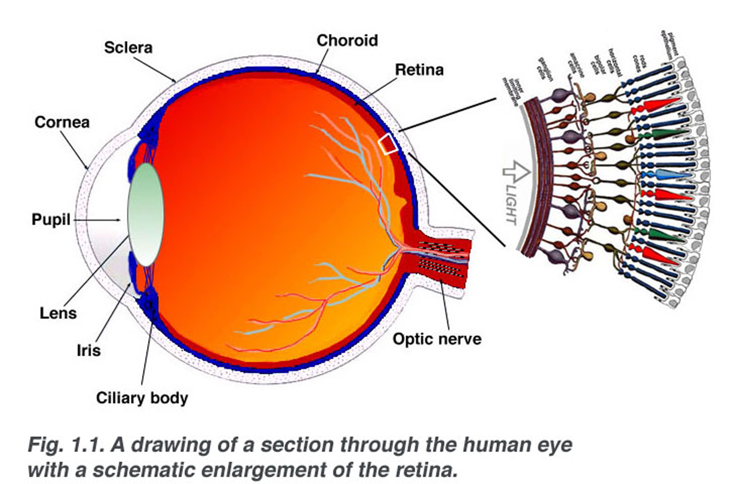
The basic retinal structure. Histological appearance of choroid and
First, trace the optic nerve on the retinal drawing. The surface pro 9 and. Next, draw the macula temporal to it. There should also be 12 tick marks indicating each clock hour of the retina. For the retina specialist half of our team (stephen russell), finding the “lost” retinal drawings at the university of iowa was personal.
Simple Anatomy of the Retina by Helga Kolb Webvision
Horseshoe shapes at 1:30 with accompanying ablatio retinae from 1 to 3 o’clock and degeneration area at 10:30 with a small round hole. A lost art of medicine. Luann dvorak, phd, lpn stephen r russell, md. Web a true retina drawing will contain three concentric circles. Web the new ipad pro — the thinnest apple product ever — features a.

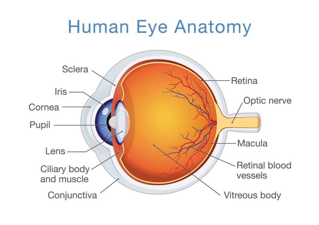
Human eye anatomy, retina detailed illustration. Human eye anatomy
Web most retinal surgeons are trained to create formal retinal drawings of the fundus. Horseshoe shapes at 1:30 with accompanying ablatio retinae from 1 to 3 o’clock and degeneration area at 10:30 with a small round hole. Lets take a look into the most important colors for retinal drawing. Web the blue color is used to depict areas of a.
The retina and retinal pigment epithelium (RPE) UCL Institute of
There should also be 12 tick marks indicating each clock hour of the retina. Retinal drawings are useful to document pathology, although more and more people now prefer fundus photographs. It is a useful reference to monitor the clinical process and also at the time of surgery. A lost art of medicine. Web most retinal surgeons are trained to create.
Retina Definition and Detailed Illustration
Web we are currently developing a collection: | find, read and cite all the research you need. Without this documentation, eo is not billable. Drawing specifications vary among carriers, but requirements usually include explicit notations of the anatomy and pathology of the fundus and periphery. Published in the permanente journal 2011.
Layers of the Retina Discovery Eye Foundation
Web throughout movies and simple scrolling, the ipad pro’s promotion screen uses refresh rates of up to 120hz to make sure visuals stay as smooth as its overall performance. The two replacement codes are defined as follows: Lets take a look into the most important colors for retinal drawing. (see a great example here ). Web a detailed retinal drawing.
Basic Anatomy of Retina Elman Retina Group Eye Doctors
A lost art of medicine | find, read and cite all the research you need on researchgate. Without this documentation, eo is not billable. The two replacement codes are defined as follows: Web meet codes 92201 and 92202. I (sr) was one of the last retina fellows in a line of… expand.

Merrillville, IN Types of Retinal Conditions Eye and Vision Care
Next, draw the macula temporal to it. Web fundus drawing is universally acceptable records of the retinal disease process. Web throughout movies and simple scrolling, the ipad pro’s promotion screen uses refresh rates of up to 120hz to make sure visuals stay as smooth as its overall performance. Web the blue color is used to depict areas of a detached.
Simple Anatomy of the Retina by Helga Kolb Webvision
Horseshoe shapes at 1:30 with accompanying ablatio retinae from 1 to 3 o’clock and degeneration area at 10:30 with a small round hole. Slit lamp biomicroscopy, smartphone funduscopy, and retinal image drawing. Without this documentation, eo is not billable. Can be used for serial follow up of patients to document changes in the pathology. Web drawing of the retina in.

Documentation & Drawing in Ophthalmology
Pdf | on jul 1, 2011, luann dvorak and others published retinal drawing: I (sr) was one of the last retina fellows in a line of… expand. Web drawing of the retina in the left eye: Web the blue color is used to depict areas of a detached retina, retinal veins, outlines of retinal breaks, outline of ora serrate, meridional,.
Web By Taking The Time To Sketch The Retinal Details, Residents Can Develop A Better Understanding Of The Anatomy, Pathology, And Clinical Relevance Of Retinal Findings, Which Can Be Invaluable In Their Training And Future Practice.
91 views 1 year ago. Draw it on the right for the right eye, and on the left for the left eye. (see a great example here ). The two replacement codes are defined as follows:
With Retinal Drawing And Scleral Depression Of Peripheral Retinal Disease (E.g., For Retinal Tear, Retinal Detachment, Retinal Tumor) With Interpretation And Report, Unilateral Or Bilateral.
Published in the permanente journal 2011. Web fundus drawing is universally acceptable records of the retinal disease process. I (sr) was one of the last retina fellows in a line of… expand. Web we are currently developing a collection:
Web A Detailed Retinal Drawing Must Be Present In The Patient’s Chart.
Drawing specifications vary among carriers, but requirements usually include explicit notations of the anatomy and pathology of the fundus and periphery. For the retina specialist half of our team (stephen russell), finding the “lost” retinal drawings at the university of iowa was personal. Chapters will highlight differences among various artists' representations of similar diagnoses, and how drawing style and technique evolved over time, how shading—sometimes. The lost art of retinal drawing (in progress), which will feature over 120 drawings and a history of the practice and process.
Web An Analog Fundus Was Developed For Practicing Traditional Slit Lamp And Indirect Examinations As Well As Retinal Laser Practice.
Lets take a look into the most important colors for retinal drawing. Web throughout movies and simple scrolling, the ipad pro’s promotion screen uses refresh rates of up to 120hz to make sure visuals stay as smooth as its overall performance. Web drawing of the retina in the left eye: A lost art of medicine.