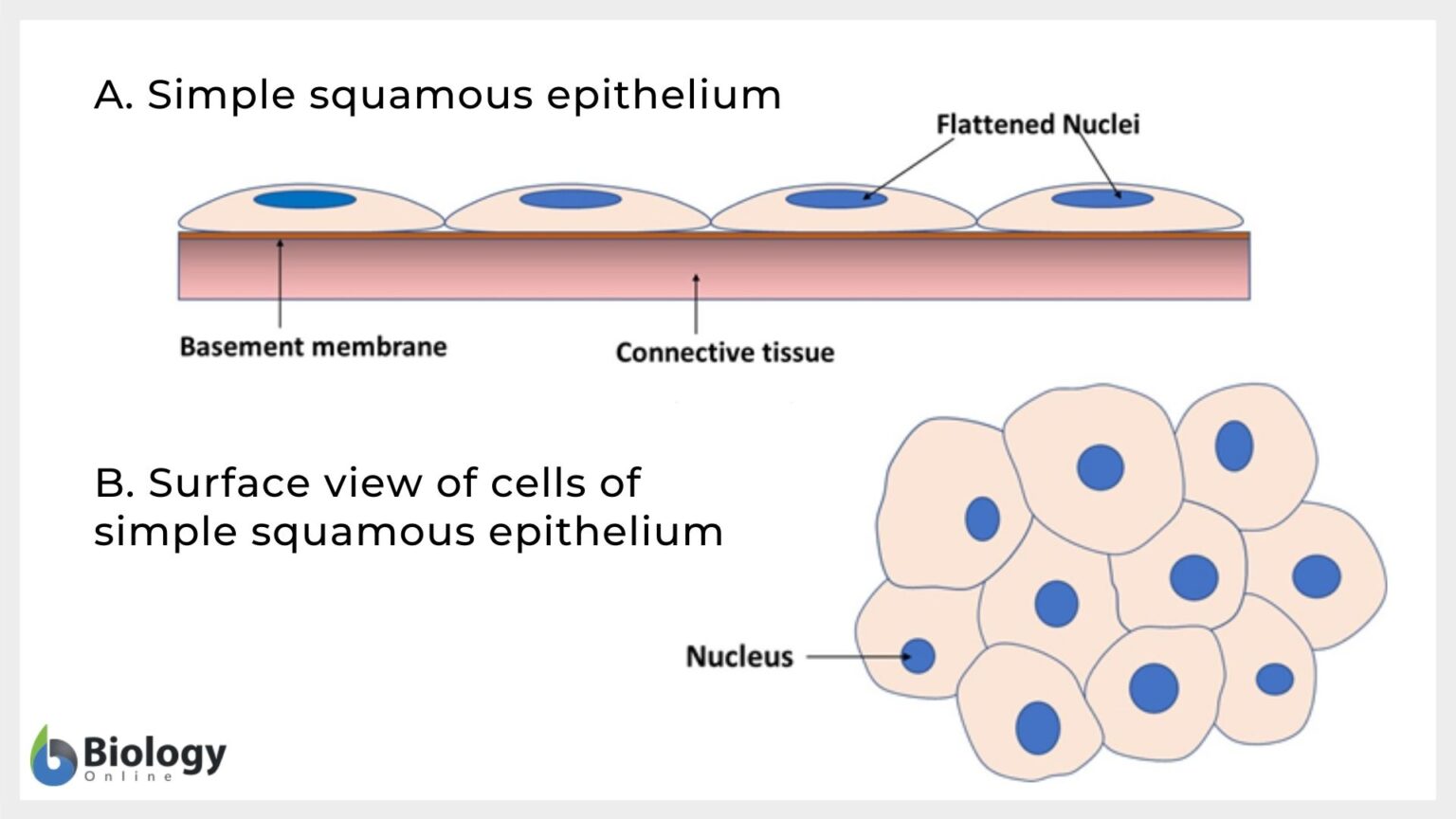
Simple Squamous Epithelium Drawing With Label
Simple Squamous Epithelium Drawing With Label - Use the image slider below to learn how to use a microscope to identify and study simple squamous epithelium in renal corpuscles of the renal (kidney) cortex. Web simple squamous epithelium consists of a single layer of flattened cells. Both surface and side view has been demonstrated in. Squamous cells are large, thin, and flat and contain a rounded nucleus. Web read more about simple squamous epithelium, 40x; Web explain the general structure and function of epithelial tissue. Web simple squamous epithelium, because of the thinness of the cell, is present where rapid passage of chemical compounds is observed. Learn about its location in the body, cells, and characteristics. Its diameter is small compared to the total diameter of the cell. Web figure 1 shows a diagram of simple squamous epithelium labeled.
The cells found in this epithelium type are flat and thin, making simple squamous epithelium ideal for lining areas where passive diffusion of gases occur. This is made up of thin, flat and hexagonal cells. The typical example of the simple squamous epithelium will be found in the lung’s alveoli, the parietal layer of the bowman’s capsule of the kidney, and the loop of henle of kidney tubules. Web in this portion, i will show you the simple squamous epithelium labeled diagrams from the different organs or parts, or structures of the animal’s body. The image can be changed using any combination of the following commands. The renal corpuscle consists of the glomerulus (capillary network derived from afferent arteriole) and bowman's capsule. Web read more about simple squamous epithelium, 40x; Distinguish between tight junctions, anchoring junctions, and gap junctions. Web simple squamous epithelium, because of the thinness of the cell, is present where rapid passage of chemical compounds is observed. Learn vocabulary, terms, and more with flashcards, games, and other study tools.
Distinguish between tight junctions, anchoring junctions, and gap junctions. Web there are three basic shapes used to classify epithelial cells. Start studying label the diagram of simple squamous epithelium. Squamous cells are large, thin, and flat and contain a rounded nucleus. Use the image slider below to learn how to use a microscope to identify and study simple squamous epithelium in renal corpuscles of the renal (kidney) cortex. Histology diagram of simple squamous epithelium histology diagram. The most obvious thing in each cell is its nucleus which is round and stained fairly darkly. Web instructor sarah phenix. Web simple squamous epithelium consists of a single layer of flattened cells. 2.6k views 3 years ago pakistan.
Simple Squamous Epithelium Labeled
Web there are three basic shapes used to classify epithelial cells. Web explain the general structure and function of epithelial tissue. Web instructor sarah phenix. The alveoli of lungs where gases diffuse, segments of kidney tubules, and the lining of capillaries are also made of simple squamous epithelial tissue. Mhs 261 common bile duct.
Simple Squamous Epithelial Tissue Under Microscope
Now you can see individual simple squamous epithelial cells (sse). Squamous cells are large, thin, and flat and contain a rounded nucleus. Web simple squamous epithelium, because of the thinness of the cell, is present where rapid passage of chemical compounds is observed. A columnar epithelial cell looks like a column or a tall rectangle. Web simple squamous epithelium consists.
Simple Squamous Epithelium Inrtroducrion , Anatomy & Function
Start studying label the diagram of simple squamous epithelium. A simple squamous epithelium, also known as pavement epithelium and tessellated epithelium, is a single layer of flattened, polygonal cells in contact with the basal lamina (one of the two layers of the basement membrane) of the epithelium. Web read more about simple squamous epithelium, 40x; A squamous epithelial cell looks.

How to draw stratified squamous epithelium easy way YouTube
Use the image slider below to learn more about the characteristics of simple squamous epithelium. The renal corpuscle consists of the glomerulus (capillary network derived from afferent arteriole) and bowman's capsule. Click on links to move to a. A simple squamous epithelium, also known as pavement epithelium and tessellated epithelium, is a single layer of flattened, polygonal cells in contact.
Simple squamous epithelium Definition and Examples Biology Online
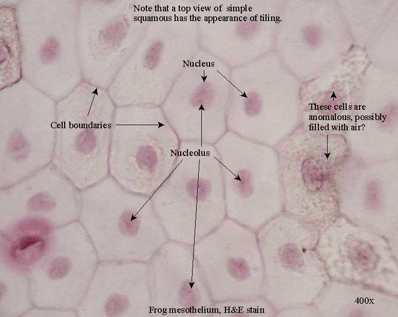
The renal corpuscle consists of the glomerulus (capillary network derived from afferent arteriole) and bowman's capsule. Mhs 224 ovary and oviduct. Learn vocabulary, terms, and more with flashcards, games, and other study tools. Web please put the total magnification in the description below your image, along with a key that defines what each label in (n= nucleus, etc.). Web simple.

Histology Image Membranous epithelium
The typical example of the simple squamous epithelium will be found in the lung’s alveoli, the parietal layer of the bowman’s capsule of the kidney, and the loop of henle of kidney tubules. 19k views 2 years ago cell biology. Web in this portion, i will show you the simple squamous epithelium labeled diagrams from the different organs or parts,.

Simple Squamous Epithelium Diagram Quizlet
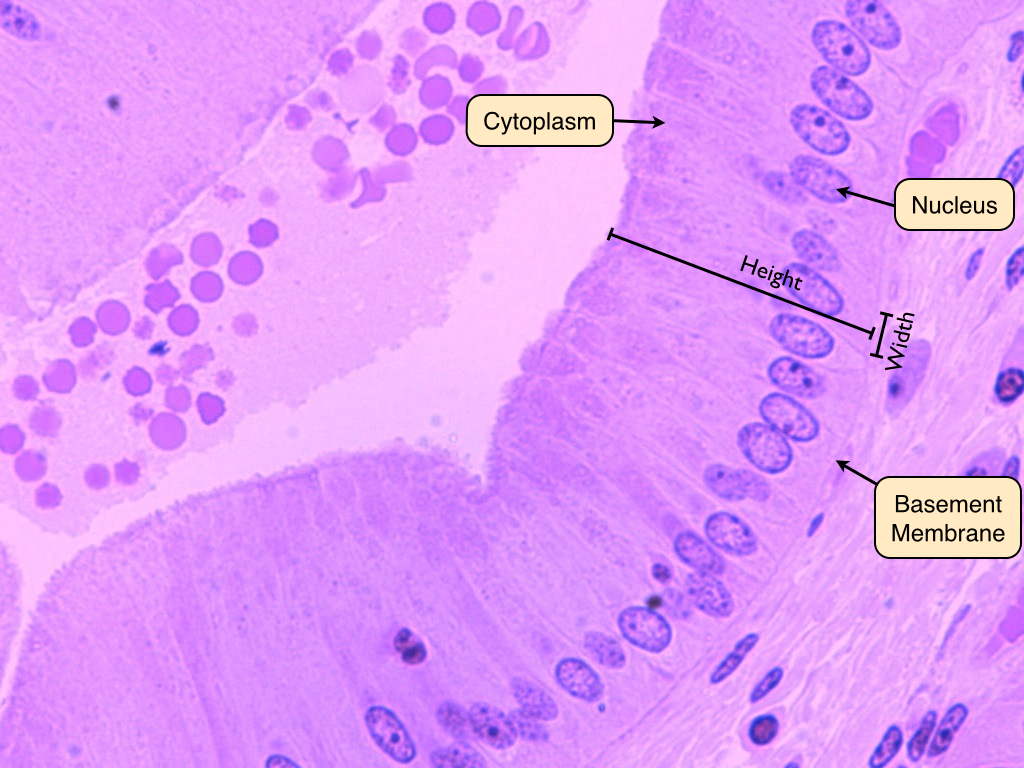
This very tall image shows almost the entire thickness of the kidney. Medical school university of minnesota minneapolis, mn. What is simple squamous epithelium? Distinguish between simple epithelia and stratified epithelia, as well as between squamous, cuboidal, and columnar epithelia. Web simple squamous epithelium, because of the thinness of the cell, is present where rapid passage of chemical compounds is.

Simple Squamous Epithelium Histology Labeled
Web simple squamous epithelium consists of a single layer of flattened cells. Web simple squmous epithelium, c.s. A squamous epithelial cell looks flat under a microscope. Its diameter is small compared to the total diameter of the cell. The thinness of these cells facilitates the transfer of materials ( e.g., gases, fluids or nutrients) across the epithelium.
Epithelial Tissue Anatomy & Physiology
Histology diagram of simple squamous epithelium histology diagram. Its diameter is small compared to the total diameter of the cell. Web in this portion, i will show you the simple squamous epithelium labeled diagrams from the different organs or parts, or structures of the animal’s body. Simple epithelium can be divided into 4 major classes, depending on the shapes of.

What is a Simple Squamous Epithelium? (with pictures)
Web in this portion, i will show you the simple squamous epithelium labeled diagrams from the different organs or parts, or structures of the animal’s body. The tissue is polarized with one surface that faces the external environment, and the other that faces the basement membrane. Squamous cells are large, thin, and flat and contain a rounded nucleus. Histology diagram.
Both Surface And Side View Has Been Demonstrated In.
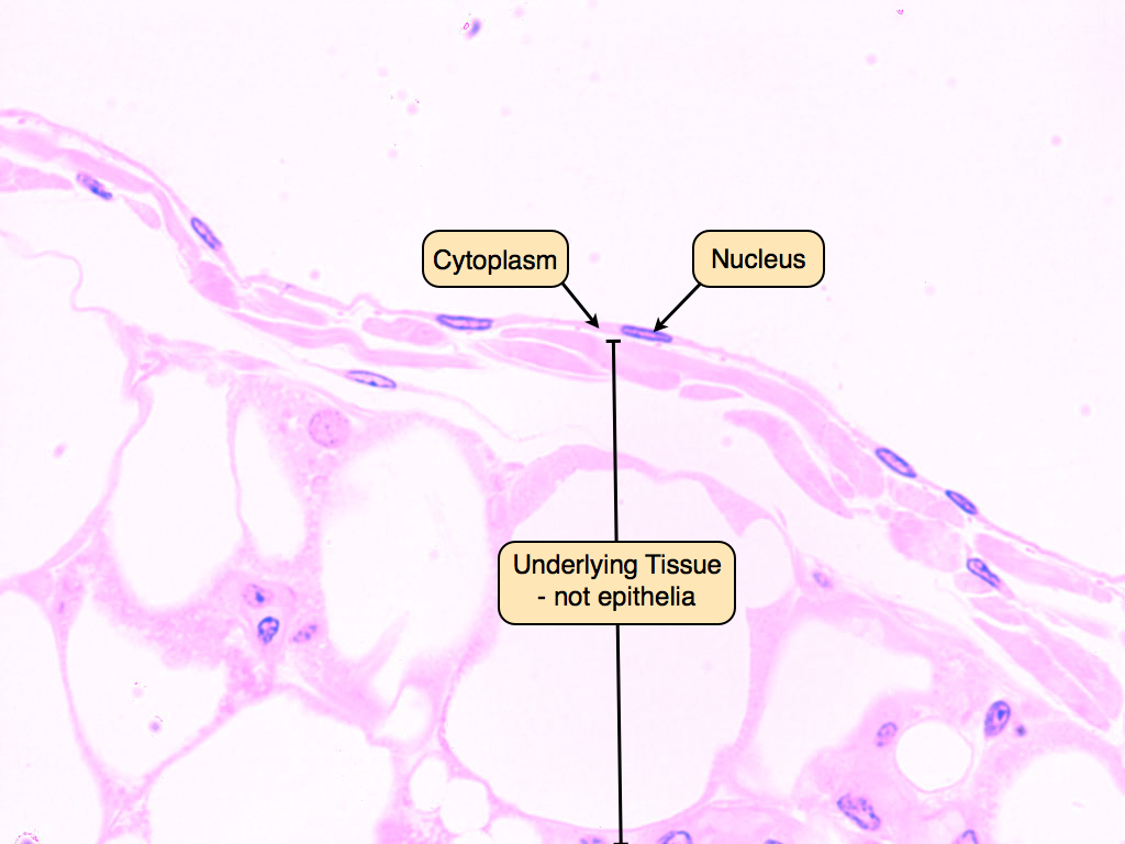
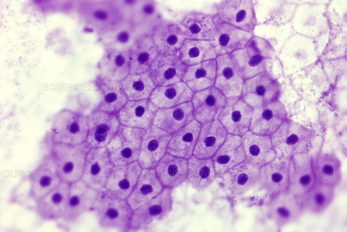
This very tall image shows almost the entire thickness of the kidney. • how to draw simple. Use the image slider below to learn how to use a microscope to identify and study simple squamous epithelium in renal corpuscles of the renal (kidney) cortex. For the sse label, try to bracket off a region of that type of epithelium, rather than just pointing to 1 cell.
The Cytoplasm Is Difficult To See Because It Is Very Thin.
Distinguish between tight junctions, anchoring junctions, and gap junctions. Each slide is shown with additional information to its right. Web simple squamous epithelium, because of the thinness of the cell, is present where rapid passage of chemical compounds is observed. Web there are three basic shapes used to classify epithelial cells.
Web Simple Squmous Epithelium, C.s.
The image can be changed using any combination of the following commands. Start studying label the diagram of simple squamous epithelium. The alveoli of lungs where gases diffuse, segments of kidney tubules, and the lining of capillaries are also made of simple squamous epithelial tissue. The thinness of these cells facilitates the transfer of materials ( e.g., gases, fluids or nutrients) across the epithelium.
See Examples And Diagrams Of Its Tissue Structure.
Now you can see individual simple squamous epithelial cells (sse). 19k views 2 years ago cell biology. The most obvious thing in each cell is its nucleus which is round and stained fairly darkly. Use the image slider below to learn more about the characteristics of simple squamous epithelium.