Smooth Muscle Tissue Drawing
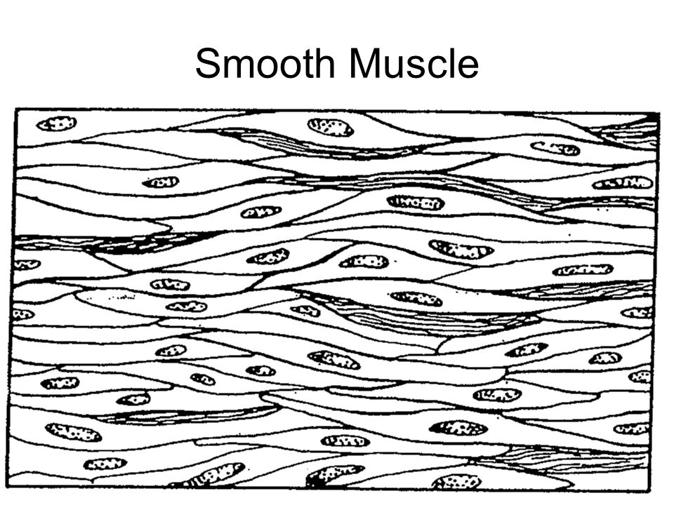
Smooth Muscle Tissue Drawing - Web smooth muscle can be confused with cardiac muscle because the cells are often running in different directions, just as they are in cardiac muscle. How to draw a muscle tissue | straight muscles | smooth muscles | cardiac muscleshello friends in this video i tell you about how to draw labelled dia. Compare this to the diagram above, and make sure you can recognise smooth muscle cells in ts and ls. Explain how smooth muscles differ from skeletal and cardiac muscles. Compare motor and sensory innervation of skeletal muscle tissue. Unlike skeletal muscle, smooth muscle is capable of maintaining tone for extended periods and often contracts involuntarily. Watch the video tutorial now. Web how to draw smooth muscle/muscle tissue diagram/how to draw smooth muscle easy.it is very easy drawing detailed method to help you.i draw the smooth muscle. It is the pen diagram of skeletal, smooth and cardiac muscle for class 10, 11 and 12. You will also find a details description of the smooth muscle fibers compared to cardiac and skeletal muscles.
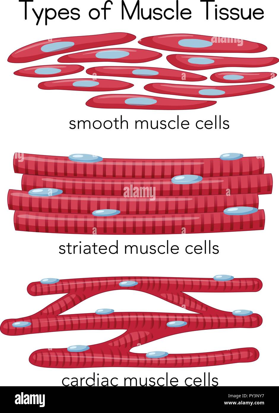
Web smooth muscle is one of three types of muscle tissue, alongside cardiac and skeletal muscle. Unlike skeletal muscle, smooth muscle is capable of maintaining tone for extended periods and often contracts involuntarily. Smooth muscle cells are a lot smaller than cardiac muscle cells, and they do not branch or connect end to end the way cardiac cells do. Watch the video tutorial now. Smooth muscle is composed of sheets or strands of smooth muscle cells. Web correlate the microscopic organization of a muscle fiber with the mechanism of contraction and relaxation. Describe the histological organization of cardiac muscle. Web in this video i have shown the simplest way of drawing muscle drawing. By the end of this section, you will be able to: Web explain the process of smooth muscle contraction.
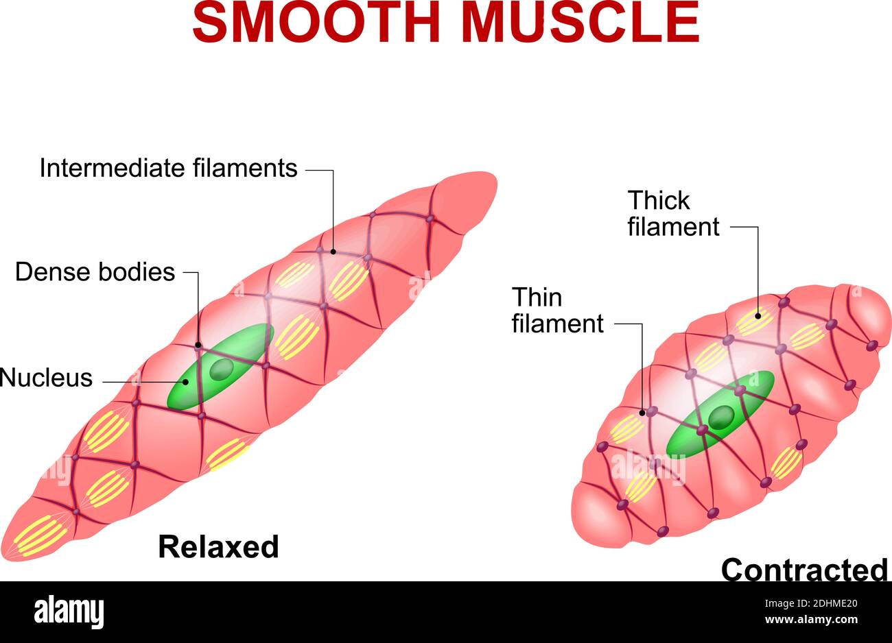
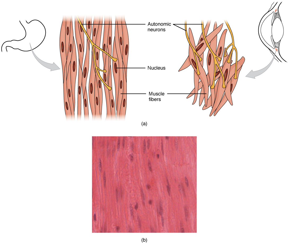
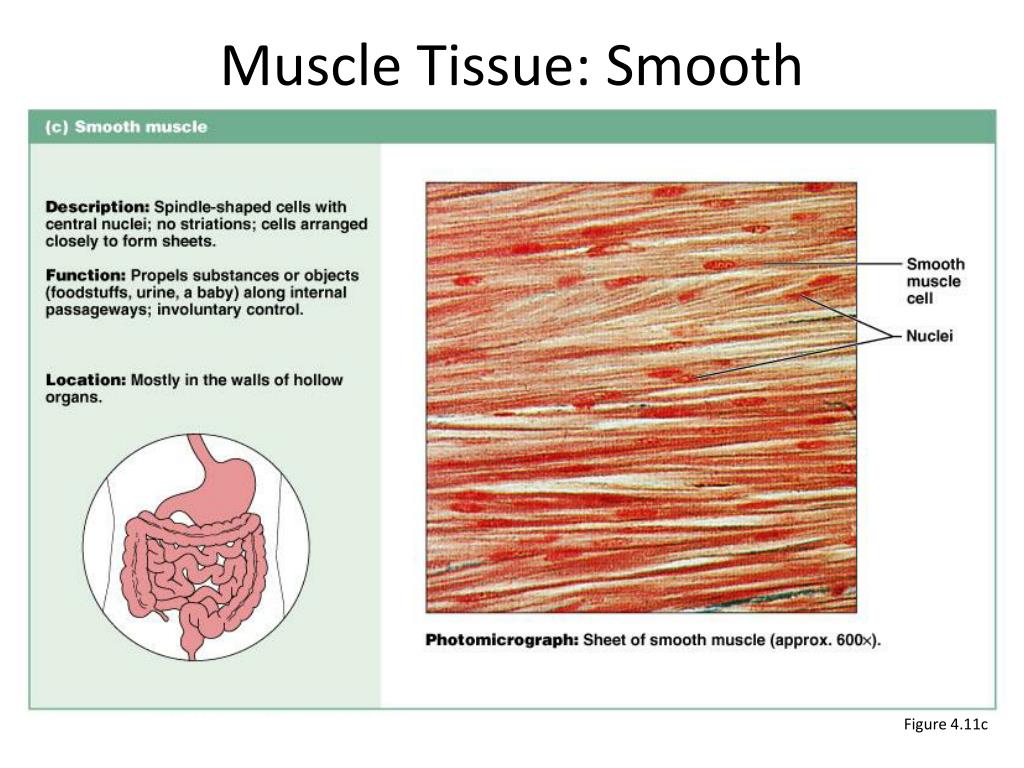
Smooth muscle cells are a lot smaller than cardiac muscle cells, and they do not branch or connect end to end the way cardiac cells do. In relaxed smooth muscle, the nuclei are elongated with rounded ends. Wall of organs like the stomach, oesophagus and intestine. Unlike cardiac and skeletal muscle cells, smooth muscle cells do not exhibit striations since their actin and myosin (thin and thick) protein filaments are not organized as sarcomeres. The area inside the box is enlarged in the next image. Smooth muscle differs from skeletal muscle in function. Web figure 10.23 smooth muscle tissue smooth muscle tissue is found around organs in the digestive, respiratory, reproductive tracts and the iris of the eye. Web smooth muscle is one of three types of muscle tissue, alongside cardiac and skeletal muscle. By the end of this section, you will be able to: Describe the histological organization of smooth muscle.

Smooth Muscle Tissue Diagram Quizlet
Web smooth muscle tissue, highlighting the inner circular layer (nuclei then rest of cells in pink), outer longitudinal layer (nuclei then rest of cells), then the serous membrane facing the lumen of the peritoneal cavity. The area inside the box is enlarged in the next image. Web smooth muscle is one of three types of muscle tissue, alongside cardiac and.
Smooth Muscle Diagram / Muscle cell diagram Smooth muscle anatomy
Web smooth muscle tissue, highlighting the inner circular layer (nuclei then rest of cells in pink), outer longitudinal layer (nuclei then rest of cells), then the serous membrane facing the lumen of the peritoneal cavity. They form the major contractile tissues of various organs. Web in this article, we'll go through the structure, function, location, characteristics, diagrams and examples of.
Smooth muscle tissue. Anatomy of a relaxed and contracted smooth muscle
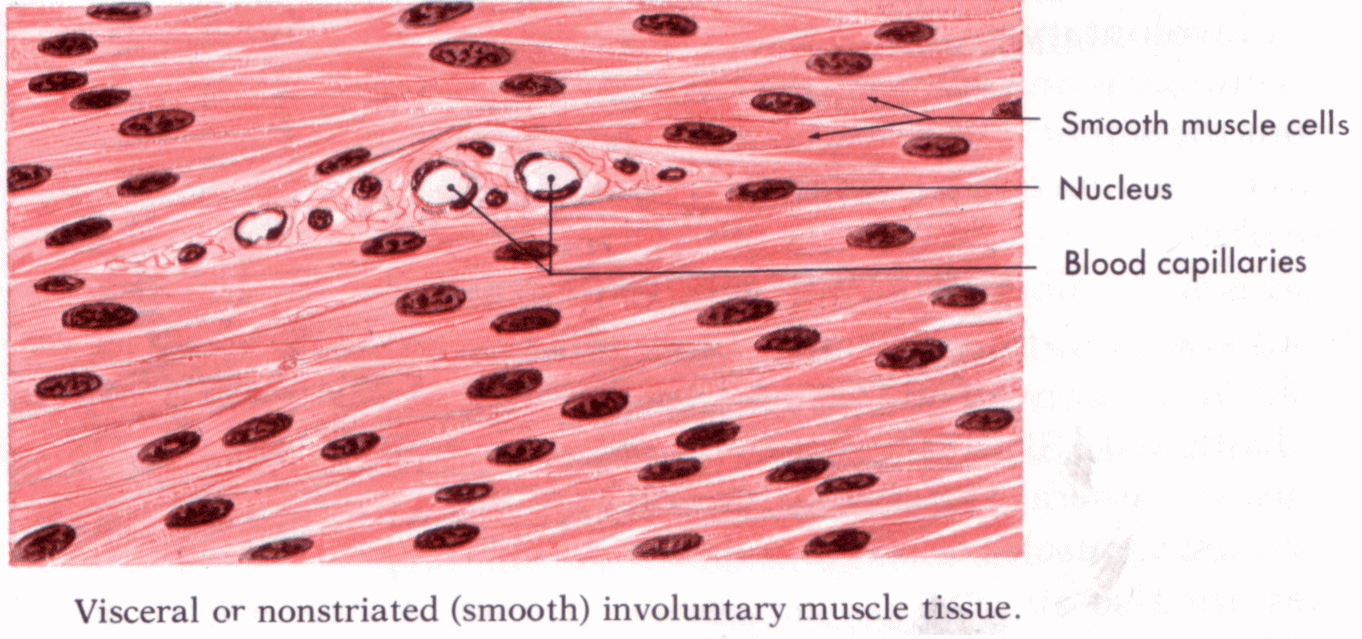
Smooth muscle differs from skeletal muscle in function. Describe the histological organization of smooth muscle. The type of muscle tissue found in the walls of blood vessels and hollow internal organs, such as the stomach, intestine etc. Web correlate the microscopic organization of a muscle fiber with the mechanism of contraction and relaxation. How to draw a muscle tissue| straight.

LM of a section through human smooth muscle tissue Stock Image P154
Web in this video i have shown the simplest way of drawing muscle drawing. Web in this simple guide, i will show you the important identifying features of the smooth muscle fibers at a light microscope with the labeled diagram. Describe the histological organization of cardiac muscle. You will also find a details description of the smooth muscle fibers compared.

Smooth Muscle Tissue Diagram Drawing ezildaricci
Explain how smooth muscles differ from skeletal and cardiac muscles. Explain how smooth muscle works with internal organs and passageways through the body. Describe the histological organization of smooth muscle. Web smooth muscle can be confused with cardiac muscle because the cells are often running in different directions, just as they are in cardiac muscle. Web correlate the microscopic organization.
Smooth Muscle Diagram Drawing Smooth Muscle Structure Function
Explain how smooth muscle differs from skeletal muscle. These muscles are found in almost all organs in the form of bundles or sheaths. How to draw a muscle. Smooth muscle is composed of sheets or strands of smooth muscle cells. Web smooth muscle is one of three types of muscle tissue, alongside cardiac and skeletal muscle.
Smooth Muscle Tissue Diagram Drawing ezildaricci
Smooth muscle is a type of muscle tissue which is used by various systems to apply pressure to vessels and organs. Web in this video i have shown the simplest way of drawing muscle drawing. The goal of this lab is to learn how to identify and describe the organization and key structural features of smooth and skeletal muscle in.
10.8 Smooth Muscle Douglas College Human Anatomy and Physiology I
Smooth muscle cells are a lot smaller than cardiac muscle cells, and they do not branch or connect end to end the way cardiac cells do. How to draw a muscle. 80k views 2 years ago class 9 diagram. Compare this to the diagram above, and make sure you can recognise smooth muscle cells in ts and ls. You will.
Smooth Muscle Diagram Drawing Notez On Nursing.... Tissues Muscle
Compare this to the diagram above, and make sure you can recognise smooth muscle cells in ts and ls. Smooth muscle is a type of muscle tissue which is used by various systems to apply pressure to vessels and organs. How to draw a muscle. Explain how smooth muscle differs from skeletal muscle. How to draw a muscle tissue| straight.
PPT Muscle Tissue PowerPoint Presentation, free download ID2093025
Describe the histological organization of cardiac muscle. It is found in numerous bodily systems, including the ophthalmic, reproductive, respiratory and gastrointestinal systems, where it functions to contract. Describe the histological organization of smooth muscle. In relaxed smooth muscle, the nuclei are elongated with rounded ends. The goal of this lab is to learn how to identify and describe the organization.
Junquiera's Basic Histology, Ch 10:
It is found in numerous bodily systems, including the ophthalmic, reproductive, respiratory and gastrointestinal systems, where it functions to contract. Web smooth muscle is one of three types of muscle tissue, alongside cardiac and skeletal muscle. Web smooth muscle can be confused with cardiac muscle because the cells are often running in different directions, just as they are in cardiac muscle. Explain how smooth muscle works with internal organs and passageways through the body.
In Relaxed Smooth Muscle, The Nuclei Are Elongated With Rounded Ends.
Web this diagram shows a few of the cells that can be seen in the stained section below. By the end of this section, you will be able to: Explain how smooth muscle differs from skeletal muscle. Web in this video i have shown the simplest way of drawing muscle drawing.
Describe The Histological Organization Of Cardiac Muscle.
Compare this to the diagram above, and make sure you can recognise smooth muscle cells in ts and ls. Smooth muscle is composed of sheets or strands of smooth muscle cells. Unlike skeletal muscle, smooth muscle is capable of maintaining tone for extended periods and often contracts involuntarily. Web explain the process of smooth muscle contraction.
Watch The Video Tutorial Now.
Web smooth muscle tissue, highlighting the inner circular layer (nuclei then rest of cells in pink), outer longitudinal layer (nuclei then rest of cells), then the serous membrane facing the lumen of the peritoneal cavity. Smooth muscle cells are a lot smaller than cardiac muscle cells, and they do not branch or connect end to end the way cardiac cells do. Web how to draw smooth muscle/muscle tissue diagram/how to draw smooth muscle easy.it is very easy drawing detailed method to help you.i draw the smooth muscle. Describe the histological organization of smooth muscle.