Spinal Cord And Spinal Nerves Review Sheet Exercise 15
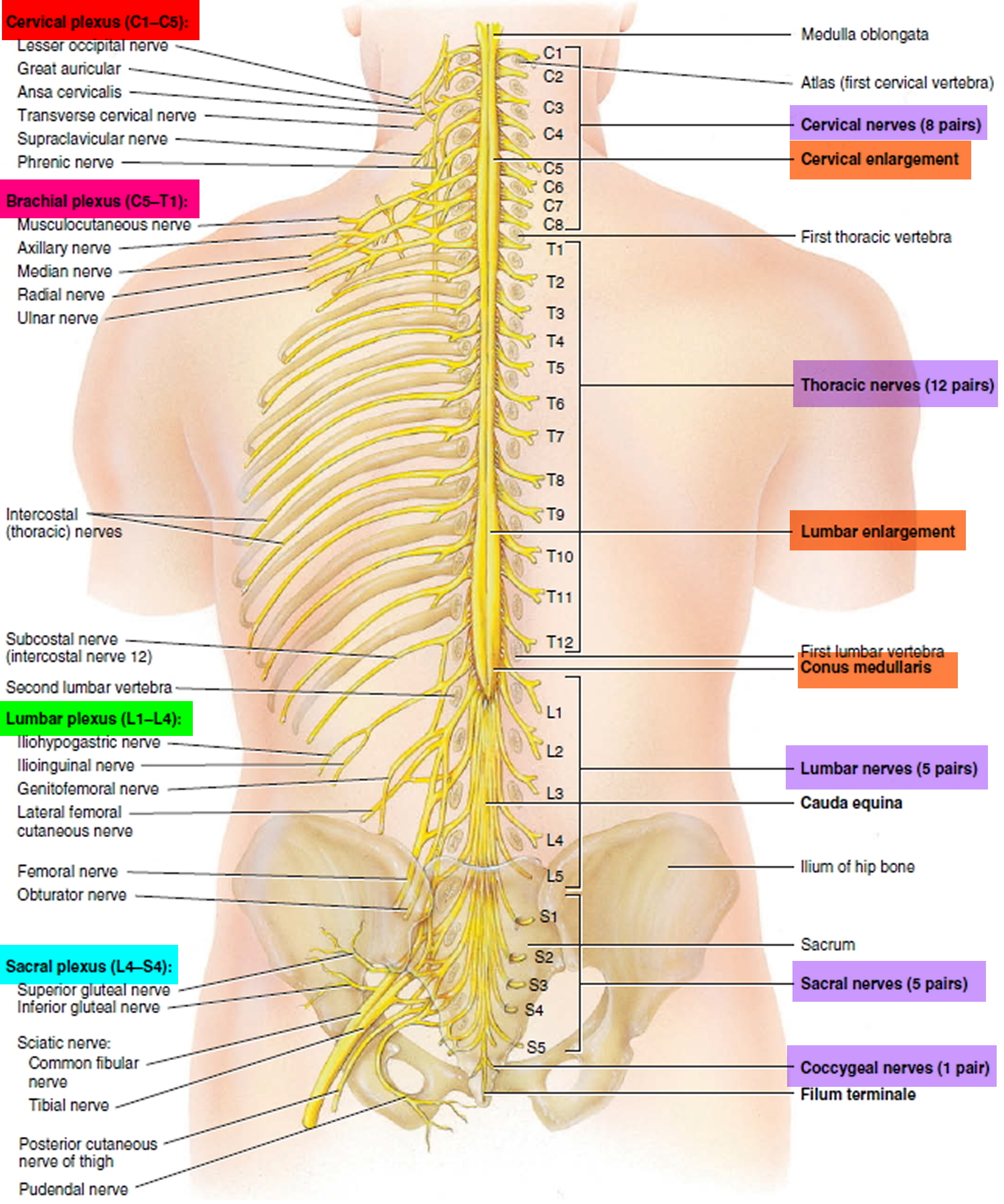
Spinal Cord And Spinal Nerves Review Sheet Exercise 15 - Web medial side of the hand. Most superior boundary of the spinal cord ________________ 2. A functional assessment of the spinal nerves that form the lumbar plexus indicated a functional loss of spinal nerve l on the right side. Study with quizlet and memorize flashcards containing terms like match. A pelvic splanchnic nerve contains. Web compare and contrast the meninges of the spinal cord and the brain. Lab exercise review sheet #2. Both the meninges of the spinal cord and the meninges of the brain contain connective tissue that make the meninges, surround and protect each. Meningeal extension beyond the spinal. Web the following peripheral nerves are derived from the lumbar plexus:
Web compare and contrast the meninges of the spinal cord and the brain. Most superior boundary of the spinal cord ________________ 2. Study with quizlet and memorize flashcards containing terms like match. Meningeal extension beyond the spinal. A pelvic splanchnic nerve contains. Both the meninges of the spinal cord and the meninges of the brain contain connective tissue that make the meninges, surround and protect each. Lab exercise review sheet #2. Web medial side of the hand. A functional assessment of the spinal nerves that form the lumbar plexus indicated a functional loss of spinal nerve l on the right side. Web the following peripheral nerves are derived from the lumbar plexus:
Most superior boundary of the spinal cord ________________ 2. Lab exercise review sheet #2. Both the meninges of the spinal cord and the meninges of the brain contain connective tissue that make the meninges, surround and protect each. Study with quizlet and memorize flashcards containing terms like match. Web the following peripheral nerves are derived from the lumbar plexus: Web compare and contrast the meninges of the spinal cord and the brain. Web medial side of the hand. Meningeal extension beyond the spinal. A functional assessment of the spinal nerves that form the lumbar plexus indicated a functional loss of spinal nerve l on the right side. A pelvic splanchnic nerve contains.
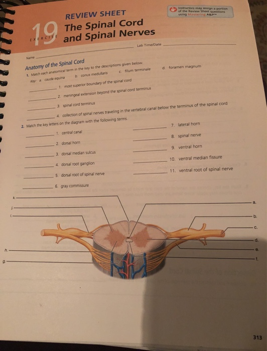
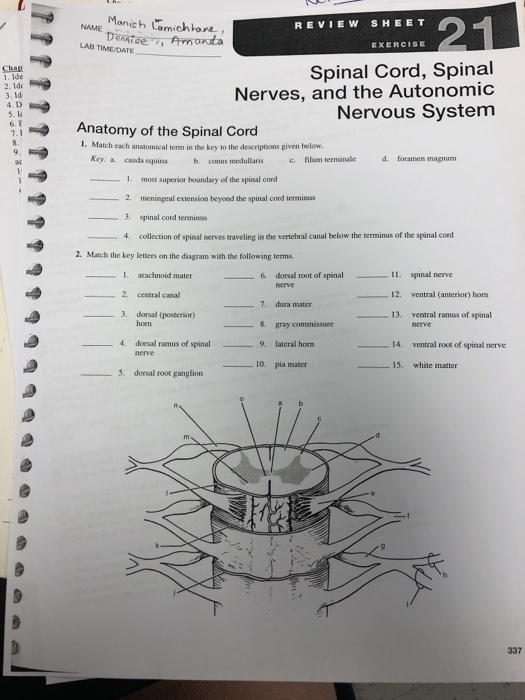
🌱 Most superior boundary of the spinal cord. Spinal Cord. 20221030
Study with quizlet and memorize flashcards containing terms like match. Most superior boundary of the spinal cord ________________ 2. Meningeal extension beyond the spinal. A functional assessment of the spinal nerves that form the lumbar plexus indicated a functional loss of spinal nerve l on the right side. A pelvic splanchnic nerve contains.
Exercise 15 Spinal Cord And Spinal Nerves Answer Key Exercise Poster
Most superior boundary of the spinal cord ________________ 2. Both the meninges of the spinal cord and the meninges of the brain contain connective tissue that make the meninges, surround and protect each. Web the following peripheral nerves are derived from the lumbar plexus: Lab exercise review sheet #2. Meningeal extension beyond the spinal.
Babinski reflex test & causes of positive Babinski reflex in adults
A pelvic splanchnic nerve contains. Web the following peripheral nerves are derived from the lumbar plexus: Lab exercise review sheet #2. Most superior boundary of the spinal cord ________________ 2. Web medial side of the hand.
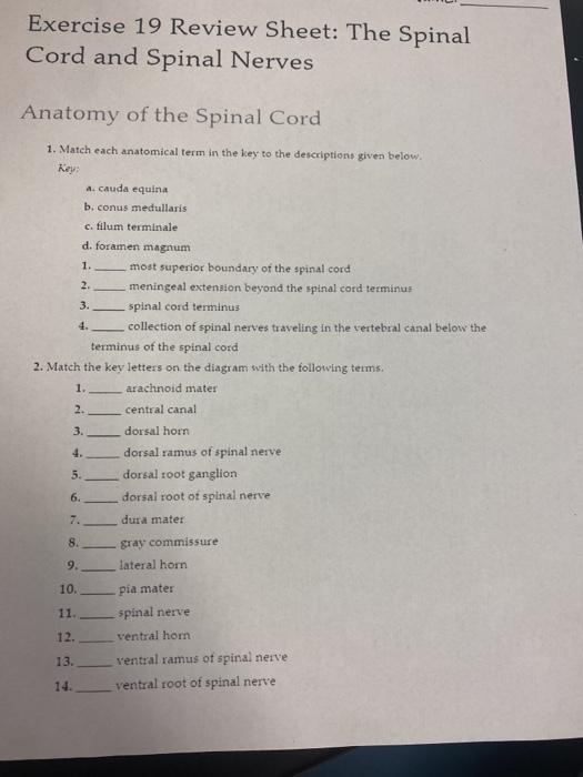
Solved Exercise 19 Review Sheet The Spinal Cord and Spinal
Meningeal extension beyond the spinal. Both the meninges of the spinal cord and the meninges of the brain contain connective tissue that make the meninges, surround and protect each. Web medial side of the hand. A pelvic splanchnic nerve contains. A functional assessment of the spinal nerves that form the lumbar plexus indicated a functional loss of spinal nerve l.

SOLUTION spinal cord spinal nerves and somatic reflexes presentation
Meningeal extension beyond the spinal. A functional assessment of the spinal nerves that form the lumbar plexus indicated a functional loss of spinal nerve l on the right side. Lab exercise review sheet #2. Study with quizlet and memorize flashcards containing terms like match. Web compare and contrast the meninges of the spinal cord and the brain.
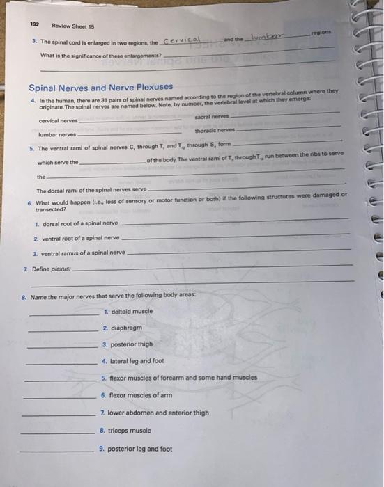
Solved 192 Review Sheet 15 regions 2. The spinal cord is
A pelvic splanchnic nerve contains. Web the following peripheral nerves are derived from the lumbar plexus: Meningeal extension beyond the spinal. Web compare and contrast the meninges of the spinal cord and the brain. Both the meninges of the spinal cord and the meninges of the brain contain connective tissue that make the meninges, surround and protect each.
Spinal Nerves What They Are and What They Do Total Community Care
Most superior boundary of the spinal cord ________________ 2. A functional assessment of the spinal nerves that form the lumbar plexus indicated a functional loss of spinal nerve l on the right side. Lab exercise review sheet #2. Both the meninges of the spinal cord and the meninges of the brain contain connective tissue that make the meninges, surround and.
Cervical Nerves
Meningeal extension beyond the spinal. A pelvic splanchnic nerve contains. Web compare and contrast the meninges of the spinal cord and the brain. Web medial side of the hand. Both the meninges of the spinal cord and the meninges of the brain contain connective tissue that make the meninges, surround and protect each.

Fillable Online Spinal cord and spinal nerves review sheet 15 answers
Most superior boundary of the spinal cord ________________ 2. Web compare and contrast the meninges of the spinal cord and the brain. Web the following peripheral nerves are derived from the lumbar plexus: A pelvic splanchnic nerve contains. Web medial side of the hand.
Simple Spinal Nerves Diagram
Web the following peripheral nerves are derived from the lumbar plexus: Study with quizlet and memorize flashcards containing terms like match. A functional assessment of the spinal nerves that form the lumbar plexus indicated a functional loss of spinal nerve l on the right side. Lab exercise review sheet #2. Web compare and contrast the meninges of the spinal cord.
A Functional Assessment Of The Spinal Nerves That Form The Lumbar Plexus Indicated A Functional Loss Of Spinal Nerve L On The Right Side.
Lab exercise review sheet #2. Web medial side of the hand. A pelvic splanchnic nerve contains. Web compare and contrast the meninges of the spinal cord and the brain.
Both The Meninges Of The Spinal Cord And The Meninges Of The Brain Contain Connective Tissue That Make The Meninges, Surround And Protect Each.
Meningeal extension beyond the spinal. Most superior boundary of the spinal cord ________________ 2. Web the following peripheral nerves are derived from the lumbar plexus: Study with quizlet and memorize flashcards containing terms like match.