Thalamus Drawing
Thalamus Drawing - Next, look at a diagram of the thalamus, and. Nerve fibers project out of the thalamus to the cerebral cortex in all directions, known as the thalamocortical radiations. From greek θάλαμος, chamber) [1] is a large mass of gray matter on the lateral walls of the third ventricle forming the dorsal part of the diencephalon (a division of the forebrain ). Web the thalamus is an ovoid, paired gray matter structure, found in the center of the brain, just superior to the brainstem. All sensory information, except for the sense of. Web do this to master the basic anatomy. Each side of the thalamus contains six groups of nuclei; This video will help you master the anatomy and function of the thalamus in a simple. Shading the bottom parts of the brain with a pen; This structure’s primary function is as a relay center through which sensory nerves transmit signals from the spinal cord and brainstem on the way to the cerebral cortex.
Web learn how to draw the thalamus and its connections with other brain structures in this neuroanatomy tutorial. Each side of the thalamus contains six groups of nuclei; Write down all the nucleus of the thalamus (just the names) on a corner of the page. Web are you looking for an easy way to draw a brain? Web the hypothalamus is a diencephalic region in the third ventricle situated caudal to the hypothalamic sulcus and the thalamus. Web the functional anatomy of the thalamus. From greek θάλαμος, chamber) [1] is a large mass of gray matter on the lateral walls of the third ventricle forming the dorsal part of the diencephalon (a division of the forebrain ). Take a piece of paper, and draw the outline of a football. Web table of contents. Oli takes us on a journey through the individual thalamic nuclei and their functions.
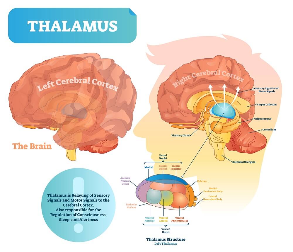
Web the hypothalamus is a diencephalic region in the third ventricle situated caudal to the hypothalamic sulcus and the thalamus. Paraventricular (midline) nuclei of thalamus. Each side of the thalamus contains six groups of nuclei; Today’s tutorial will show you how to draw a simple brain quickly and easily with just a few simple steps. Informational pathways flow into the thalamus from all areas of the central nervous system. The thalamus is a small, bilateral structure in the diencephalon that integrates signals from many areas of the cns. Its functions include relaying sensory and motor signals to the cerebral cortex and regulating consciousness, sleep, and alertness. Web the functional anatomy of the thalamus. This video will help you master the anatomy and function of the thalamus in a simple. Oli takes us on a journey through the individual thalamic nuclei and their functions.
Thalamic nuclei Connections, functions and anatomy Kenhub
The right and left thalami are connected by a mass of gray matter called the interthalamic adhesion. Web the hypothalamus is a diencephalic region in the third ventricle situated caudal to the hypothalamic sulcus and the thalamus. This video will help you master the anatomy and function of the thalamus in a simple. Each side of the thalamus contains six.

The Brain Anatomy the Thalamus Stock Illustration Illustration of
Web do this to master the basic anatomy. Web learn how to draw the thalamus and its connections with other brain structures in this neuroanatomy tutorial. Its functions include relaying sensory and motor signals to the cerebral cortex and regulating consciousness, sleep, and alertness. Web the drawing shows some areas of the brain associated with different parts of the body..
Thalamus The Definitive Guide Biology Dictionary
A growing number of nuclei isolated but. Web the thalamus, or the dorsal and ventral thalamus collectively, are two oval structures made up of gray matter at the base of the cerebrum. Web the thalamus (derived from the greek meaning “inner chamber”) is a midline symmetrical structure within the brain, situated between the cerebral cortex and midbrain. Shading the bottom.

3d rendered medically accurate illustration of the thalamus Insights
As you can see, larger areas of the brain in this region are associated with the hands, face, and tongue than the legs and feet. All sensory information, except for the sense of. Web are you looking for an easy way to draw a brain? Nerve fibers project out of the thalamus to the cerebral cortex in all directions, known.

Thalamus brain drawing Stock Photo Alamy
Web the thalamus (derived from the greek meaning “inner chamber”) is a midline symmetrical structure within the brain, situated between the cerebral cortex and midbrain. Web the thalamus ( pl.: It’s known as a relay station of all incoming motor (movement) and sensory information — hearing, taste, sight and touch. Today’s tutorial will show you how to draw a simple.

Anatomy Of Thalamus
Forming the brain with a pencil sketch; Web what is the thalamus? Today’s tutorial will show you how to draw a simple brain quickly and easily with just a few simple steps. Nerve fibers project out of the thalamus to the cerebral cortex in all directions, known as the thalamocortical radiations. It’s known as a relay station of all incoming.

Thalamus Neurology Medbullets Step 1
It’s known as a relay station of all incoming motor (movement) and sensory information — hearing, taste, sight and touch. A growing number of nuclei isolated but. As you can see, larger areas of the brain in this region are associated with the hands, face, and tongue than the legs and feet. Web after printing invention (∼1450), macroscopic drawings of.
Thalamus Anatomy and Blood Supply Kenhub
As you can see, larger areas of the brain in this region are associated with the hands, face, and tongue than the legs and feet. Its functions include relaying sensory and motor signals to the cerebral cortex and regulating consciousness, sleep, and alertness. It’s known as a relay station of all incoming motor (movement) and sensory information — hearing, taste,.

Thalamus The Science of Psychotherapy
Its functions include relaying sensory and motor signals to the cerebral cortex and regulating consciousness, sleep, and alertness. Outlining intricate parts of the brain; A growing number of nuclei isolated but. Oli takes us on a journey through the individual thalamic nuclei and their functions. Web the thalamus ( pl.:

Neuroanatomy Glossary Thalamus Draw It to Know It
Web the thalamus is an ovoid, paired gray matter structure, found in the center of the brain, just superior to the brainstem. Web the diencephalon is represented by a posterior view of the thalamus with its nuclei, followed by a sagittal section to locate the hypothalamus (including mammillary bodies, tuber cinereum, lamina terminalis), the epithalamus (with the pineal gland and.
Web The Functional Anatomy Of The Thalamus.
Web the thalamus, or the dorsal and ventral thalamus collectively, are two oval structures made up of gray matter at the base of the cerebrum. Web the thalamus ( pl.: Web the hypothalamus is a diencephalic region in the third ventricle situated caudal to the hypothalamic sulcus and the thalamus. Web do this to master the basic anatomy.
Take A Piece Of Paper, And Draw The Outline Of A Football.
Write down all the nucleus of the thalamus (just the names) on a corner of the page. Web after printing invention (∼1450), macroscopic drawings of thalamus become splendid. Next, look at a diagram of the thalamus, and. Web are you looking for an easy way to draw a brain?
Informational Pathways Flow Into The Thalamus From All Areas Of The Central Nervous System.
It is involved in sexual arousal, emotional response, endocrine regulation, sexual development, thermoregulation, regulation of satiety and hunger, and is also involved in osmoregulation. This video will help you master the anatomy and function of the thalamus in a simple. Web in this chapter, we will draw the nuclei of the thalamus. Additionally, the thalamus plays a role in alertness, sleep, and consciousness as well as learning and memory.
Paraventricular (Midline) Nuclei Of Thalamus.
Outlining intricate parts of the brain; The thalamus is made up of two symmetrical structures formed from the diencephalon. Web table of contents. The right and left thalami are connected by a mass of gray matter called the interthalamic adhesion.