Transitional Epithelium Drawing
Transitional Epithelium Drawing - Web i have described how to make transitional epithelium.this is the last video of epithelium.now i will upload how to draw connective tissue.plz share, li. Web dr naveed anjum. Web transitional epithelium is a stratified tissue made of multiple cell layers, where the cells constituting the tissue can change shape depending on the distention in. Useful for all medical students. Drawn by using h & e pencils. Single layer of cuboidal shaped cells. We will make it easy for medical students to make histology diagrams in short time during practical. Web description and photographs of transitional epithelium in the kidney and bladder, including electron micrographs showing distensible surface cells. Web transitional epithelium is a type of stratified epithelium composed of several layers of cells, with the morphology of cells varying depending on the function of the organ. It rapidly adapts to distention and contraction by changing from a.
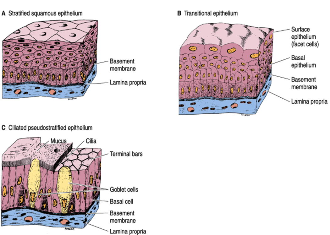
Web i have described how to make transitional epithelium.this is the last video of epithelium.now i will upload how to draw connective tissue.plz share, li. See images, diagrams, and interactive activities to identify and. Drawn by using h & e pencils. The transitional epithelium usually appears cuboidal when relaxed and squamous when stretched. Web learn about the structure and function of transitional epithelium, a stratified tissue that lines the urinary tract. Web transitional epithelium is a stratified tissue made of multiple cell layers, where the cells constituting the tissue can change shape depending on the distention in. Transitional epithelium is a type of stratified epithelium. Web description and photographs of transitional epithelium in the kidney and bladder, including electron micrographs showing distensible surface cells. The image shows the wall. Web drawing histological diagram of transitional epithelia.
Web transitional epithelium (urothelium) is a specialized stratified epithelium found in the lower urinary tract. This tissue consists of multiple layers of epithelial cells which can contract and expand in order to adapt to the degree. Transitional epithelium is a type of tissue that changes shape in response to stretching (stretchable epithelium). Drawn by using h & e pencils. See images, diagrams, and interactive activities to identify and. The transitional epithelium usually appears cuboidal when relaxed and squamous when stretched. Transitional epithelium is a type of stratified epithelium. Web learn to draw transitional epithelium histology diagram ( for medical students) The urothelium or epithelium of transition lines your urinary bladder ureters. Web dr naveed anjum.

Transitional Epithelium Diagram Quizlet
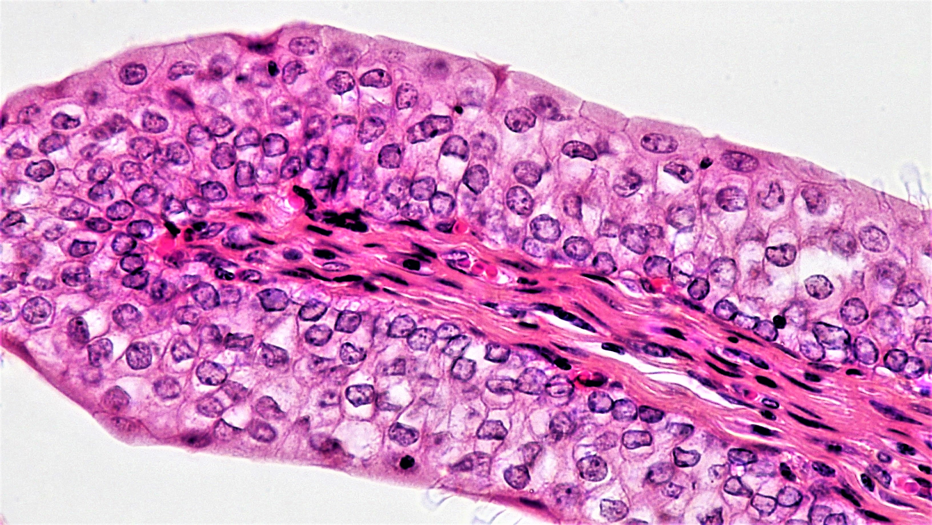
Transitional epithelium is a type of tissue that changes shape in response to stretching (stretchable epithelium). Web transitional epithelium is a type of stratified epithelium composed of several layers of cells, with the morphology of cells varying depending on the function of the organ. Web photo taken by theresa carrera transitional epithelium (400x) the blue bracket indicates the transitional epithelium.
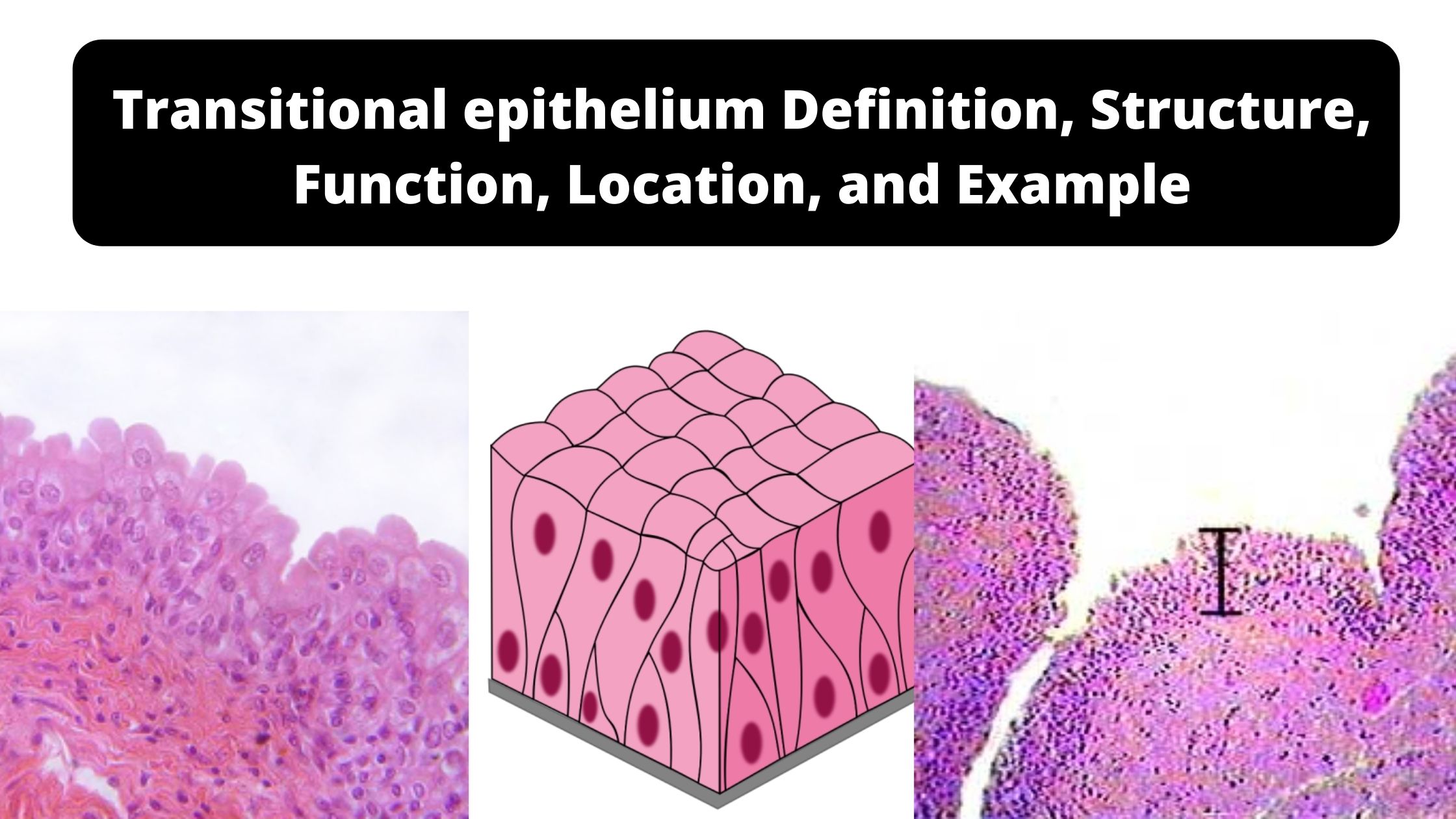
Transitional epithelium Definition, Structure, Function, Location, and
We will make it easy for medical students to make histology diagrams in short time during practical. Web i have described how to make transitional epithelium.this is the last video of epithelium.now i will upload how to draw connective tissue.plz share, li. Web transitional epithelium is a stratified tissue in which the cells are all have a fairly round shape.

Transitional epithelium Histology slides, Medical laboratory science
Web learn to draw transitional epithelium histology diagram ( for medical students) Web transitional epithelium is a type of stratified epithelium composed of several layers of cells, with the morphology of cells varying depending on the function of the organ. Drawn by using h & e pencils. This tissue consists of multiple layers of epithelial cells which can contract and.

Transitional Epithelium Function, Location & Characteristics Lesson
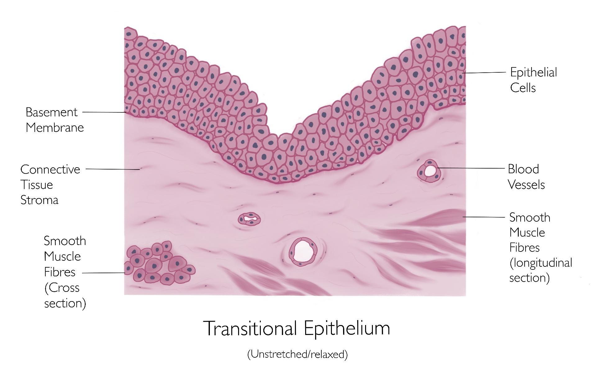
This tissue consists of multiple layers of epithelial cells which can contract and expand in order to adapt to the degree. The image shows the wall. The urothelium or epithelium of transition lines your urinary bladder ureters. Web transitional epithelium is a stratified tissue made of multiple cell layers, where the cells constituting the tissue can change shape depending on.
Transitional Epithelium
Single layer of cuboidal shaped cells. Web transitional epithelium is a layer of cells that forms the mucosal lining of your ureters, a portion of your urethra, and your urinary bladder. Web transitional epithelium is a stratified tissue in which the cells are all have a fairly round shape when the organ it lines is not distended (stretched out). Web.
MBBS Medicine (Humanity First) EPITHELIUM
The urothelium or epithelium of transition lines your urinary bladder ureters. See images, diagrams, and interactive activities to identify and. Web simple cuboidal epithelium. 323 views 2 years ago. Single layer of cuboidal shaped cells.
Simple For Transitional Epithelium Professional 2 Easily Interior
Web i have described how to make transitional epithelium.this is the last video of epithelium.now i will upload how to draw connective tissue.plz share, li. Single layer of cuboidal shaped cells. Web photo taken by theresa carrera transitional epithelium (400x) the blue bracket indicates the transitional epithelium when the bladder is contracted (not distended). Transitional epithelium is a type of.

Transitional Epithelia (urinary bladder) Human anatomy and physiology
Web learn to draw transitional epithelium histology diagram ( for medical students) Single layer of cuboidal shaped cells. Web drawing histological diagram of transitional epithelia. Web in humans, transitional epithelium (urothelium) and nonkeratinizing stratified squamous epithelium, which becomes more prominent in the distal urethra, line the urethra. The urothelium or epithelium of transition lines your urinary bladder ureters.

transitional epithelium Diagram Quizlet
Single layer of cuboidal shaped cells. Transitional epithelium is a type of tissue that changes shape in response to stretching (stretchable epithelium). Transitional epithelium is a type of stratified epithelium. Web i have described how to make transitional epithelium.this is the last video of epithelium.now i will upload how to draw connective tissue.plz share, li. On surface view, cells look.

Transitional Epithelium Diagram Quizlet
See images, diagrams, and interactive activities to identify and. Web photo taken by theresa carrera transitional epithelium (400x) the blue bracket indicates the transitional epithelium when the bladder is contracted (not distended). Useful for all medical students. Single layer of cuboidal shaped cells. We will make it easy for medical students to make histology diagrams in short time during practical.
Web Transitional Epithelium Is A Stratified Tissue In Which The Cells Are All Have A Fairly Round Shape When The Organ It Lines Is Not Distended (Stretched Out).
On surface view, cells look like mosaic (hexagonal) examples: Web in humans, transitional epithelium (urothelium) and nonkeratinizing stratified squamous epithelium, which becomes more prominent in the distal urethra, line the urethra. Transitional epithelium is a type of stratified epithelium. Web transitional epithelium is a layer of cells that forms the mucosal lining of your ureters, a portion of your urethra, and your urinary bladder.
323 Views 2 Years Ago.
Web i have described how to make transitional epithelium.this is the last video of epithelium.now i will upload how to draw connective tissue.plz share, li. Useful for all medical students. Web photo taken by theresa carrera transitional epithelium (400x) the blue bracket indicates the transitional epithelium when the bladder is contracted (not distended). Web learn about the structure and function of transitional epithelium, a stratified tissue that lines the urinary tract.
This Tissue Consists Of Multiple Layers Of Epithelial Cells Which Can Contract And Expand In Order To Adapt To The Degree.
Web transitional epithelium is a type of stratified epithelium composed of several layers of cells, with the morphology of cells varying depending on the function of the organ. The image shows the wall. Single layer of cuboidal shaped cells. Transitional epithelium is a type of tissue that changes shape in response to stretching (stretchable epithelium).
Web Transitional Epithelium Is A Stratified Tissue Made Of Multiple Cell Layers, Where The Cells Constituting The Tissue Can Change Shape Depending On The Distention In.
Web description and photographs of transitional epithelium in the kidney and bladder, including electron micrographs showing distensible surface cells. Web transitional epithelium (urothelium) is a specialized stratified epithelium found in the lower urinary tract. Web learn to draw transitional epithelium histology diagram ( for medical students) See images, diagrams, and interactive activities to identify and.